Reticular Erythematous Mucinosis or REM is a rare dermatosis characterised by papules and plaques on the upper trunk which often coalesce into a reticulated pattern. Some authors consider this entity a form of cutaneous lupus erythematosus.
Histology of reticular erythematous mucinosis
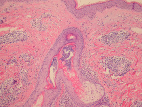
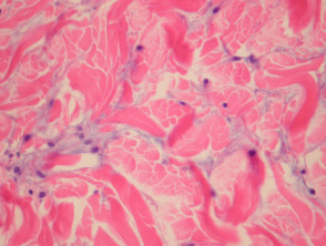
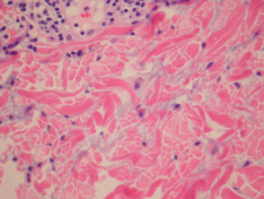
In REM, sections show a superficial and deep perivascular and periadnexal mononuclear infiltrate (figure 1). The epidermis is uninvolved. The presence of copious mucin between collagen fibres of the dermis is characteristic of the disorder (figures 2, 3).
Reticular Erythematous Mucinosis pathology
Special studies for reticular erythematous mucinosis
The mucin may be difficult to appreciate on routine H-E sections. Special stains for mucin (such as colloidal iron or hyaluronic acid) can be helpful to confirm the presence of dermal mucin.
Differential diagnosis of reticular erythematous mucinosis
Scleroedema – This entity also shows mucin deposition but typically lacks an associated inflammatory infiltrate.
Lupus – Tumid lupus can look identical to REM. The clinical presentation can help distinguish these entities. Some authorities consider REM a form of lupus.