DermNet provides Google Translate, a free machine translation service. Note that this may not provide an exact translation in all languages

Translate

See your doctor for a skin check

Author: Vanessa Ngan, Staff Writer; Copy Editor: Clare Morrison; Chief Editor: Dr Amanda Oakley, Dermatologist, Hamilton, New Zealand, October 2013. About Melanoma is sponsored by the New Zealand Dermatological Society Incorporated.


toc-icon

If you find a suspicious looking mole or you’re just not sure about a spot you find on your body, go and see your doctor. This could save your life!

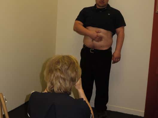
Your doctor is trained to identify suspect moles or spots as potentially melanoma. At your visit be prepared for some or all of the following things:

  • Answering questions such as whether anyone else in your family has or had skin cancer including melanoma; if you find it hard to tan; if you work outdoors; whether you stayed out in the sun a lot when you were young; if you have had bad sunburn; and, if you have noticed changes in your mole over the last few weeks.
  • Removing most of your clothes, so your doctor can do a full skin check. You may wear a gown for modesty.
  • Your doctor may use a magnifying glass with a light source or a special hand-held tool called a dermatoscope to look closely at your moles and spots.
  • Your doctor may take measurements and photographs of any suspicious moles so they can be used later on for comparison or for referral to an expert for advice.
  • Your doctor may check to see if your lymph nodes are enlarged. These are found under the skin of your armpits, around your neck, and in your groin area.
Getting a good look at your spots
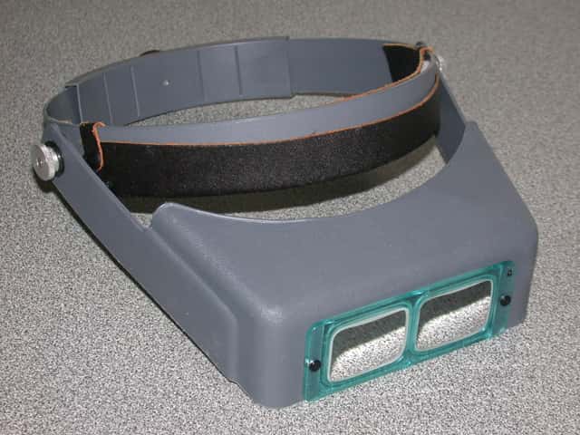
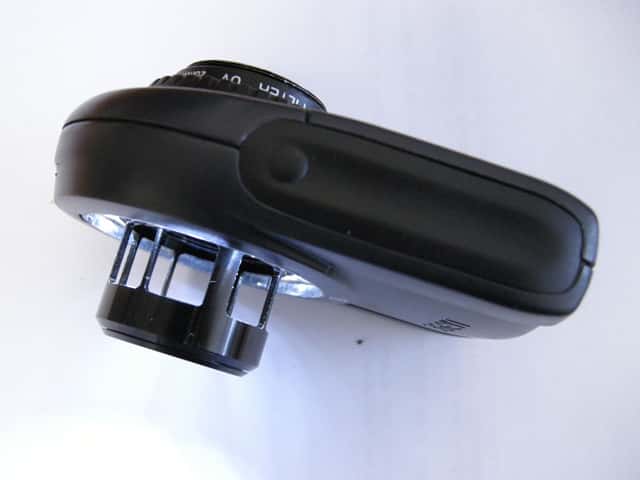
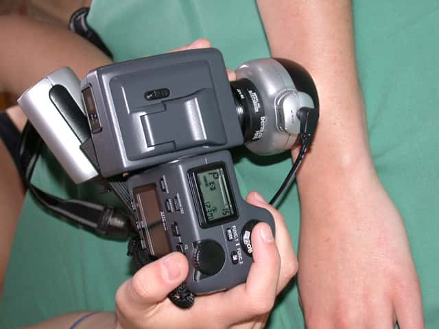
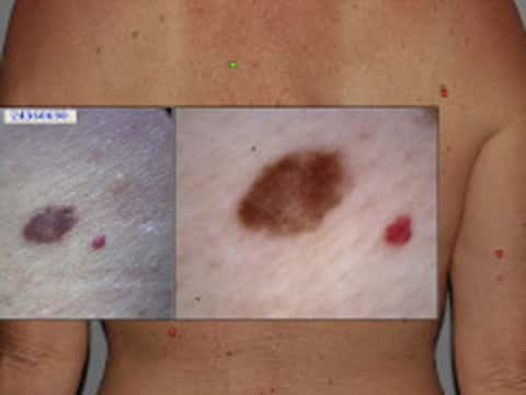
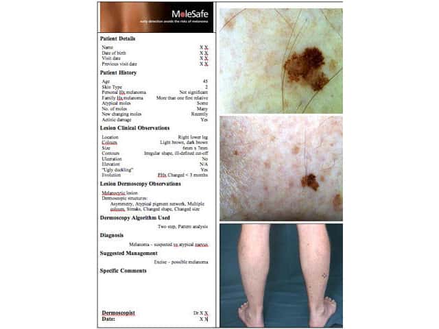
A good light, magnification and photography help identify melanoma
Optivisor Dermlite Lumio dermatoscopy Dermlite DL2 HR Pro dermatoscopy Dermatoscopic imaging

Mole mapping

For most people self skin-checks done every 1–3 months is all that is needed to pick up early signs of skin cancer and melanoma.

However, a detection programme may be useful for those at high risk of melanoma. The term ‘mole mapping’ has been used in several different ways but usually refers to a monitoring programme where:

  • High quality photographs of your whole body’s skin surface have been taken using a digital camera
  • Images are evaluated by an expert in skin cancer, usually a dermatologist
  • A report is given to you or your referring doctor which includes suspected diagnoses and recommendations for treatment
  • Your images and reports are securely stored in a database for future comparisons
  • Copies of images may be given to you to aid in self skin-checks

The mole mapping process is sometimes called teledermatoscopy or digital dermatoscopic surveillance. It is most suitable for people with many moles, especially if they have atypical moles.

The outcome of your first mole map will determine when you have your next one. A follow-up visit in 3 to 6 months may be needed to photograph a mole that is borderline to ensure it is stable.

Otherwise you may have a follow-up mole map every one to two years or at such a time as recommended by your doctor.

A less technical mole mapping procedure is your doctor simply marking spots on a cartoon drawing of a body to indicate the position of moles and spots that might be of concern. Your doctor may examine your moles using a dermatoscope and take photographs for future reference.

Mole mapping*
Taking body shots A mole that's been mapped Body shots Report for patient and doctor
Mole mapping Mole mapping Mole mapping Mole mapping

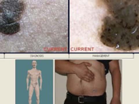
*Images supplied by MoleMap NZ

If I have a suspicious skin spot, what happens next?

If you have a suspected skin cancer, including melanoma, your doctor may arrange to remove it surgically at your usual medical centre.

If your doctor is unsure of the diagnosis, or if your suspicious mole needs the attention of an expert, you may be referred to a specialist. Skin cancer specialists include dermatologists, plastic surgeons and other highly trained doctors.


Related information

Sign up to the newsletter