What is a haematological disease?
Haematological diseases are a diverse range of conditions affecting the constituents of blood. This includes disorders of the blood cells (red cells, white cells and platelets) and cancerous conditions affecting these blood cells. Skin signs of haematological disease described here are helpful in diagnosis and may also cause complications.
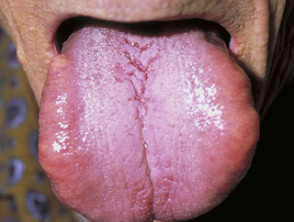
Anaemia
Nutritional anaemia
Anaemia can be the result of nutrient deficiencies, for example, iron, B12 or folate.
Skin manifestations are:
- Pallor of the conjunctiva (eyes) and palmar creases
- Glossitis (smooth red tongue)
- Poikilodermatous hypopigmentation
- Hyperpigmentation may be a sign of B12 and folate deficiencies
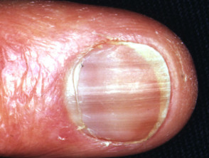
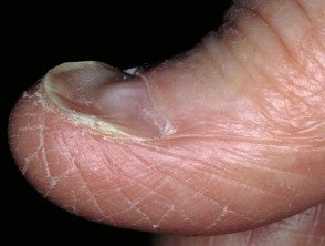
- Brittle nails and koilonychia
Skin signs of anaemia
Haemolytic anaemia
Haemolytic anaemia, for example, due to sickle cell anaemia, results from the destruction of red blood cells, which leads to additional symptoms:
- Pruritus
- Jaundice
- Petechiae and haemosiderosis (small brown macules).
Myeloproliferative disorders
The process by which blood cells are produced (haematopoiesis) gives rise to cells in either the lymphoid or myeloid lineage.
Polycythaemia
Polycythaemia vera is an example of a chronic myeloproliferative disorder of myeloid cells that results in an increased red cell mass.
Skin manifestations are:
- Plethora/ruddy cyanosis (florid complexion)
- Aquagenic pruritus (itching of skin triggered by contact with water)
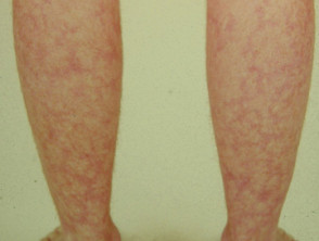
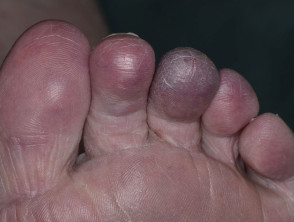
- Erythromelalgia (redness and burning pain of hands/feet)
- Livedo reticularis (purplish lace-like discolouration)
- Acrocyanosis (bluish-purplish discolouration of hands/feet)
- Pyoderma gangrenosum (ulcers).
Skin signs of myeloproliferative disease
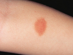
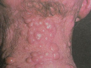
Mast cell disease
Mast cell disease also arises from a myeloid disorder in which mast cells accumulate in different organs and tissues in the body. This can be broadly divided into cutaneous mastocytosis, which is more common in children, and systemic mastocytosis, which is more common in adults.
Skin manifestations are:
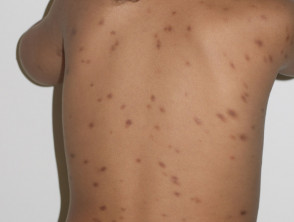
- Maculopapular cutaneous mastocytosis (tan/brown papules in children, reddish-brown macules and papules in adults). Also known as urticaria pigmentosa.
- Darier sign (erythema and urticaria when affected skin is rubbed)
- Pruritus
- Mastocytomas (rare in adults)
Skin signs of mast cell disease
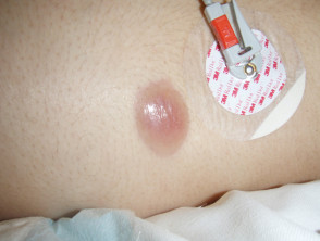
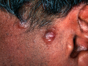
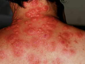
Leukaemia cutis
Leukaemia cutis is a rare manifestation of undiagnosed or previously treated myeloproliferative and lymphoproliferative disorders, eg acute myeloid leukaemia (AML), non-Hodgkin lymphoma. It is a result of infiltration of the skin by leukaemic cells, resulting in variable papules, nodules and plaques. Typically, they can be pinkish, violaceous (purplish) or darker than surrounding normal skin. In addition, they are almost always non-tender and palpable with a firm, indurated texture.
Leukaemia cutis
VEXAS syndrome
VEXAS syndrome is an acquired genetic condition seen in middle-aged men characterised by myelodysplastic haematological disorders and autoinflammatory features particularly involving the skin.
Lymphoproliferative disorders
Lymphoproliferative disorders arise from disorders of stem cells of the lymphoid lineage. Systemic forms of lymphoma, Hodgkin lymphoma and non-Hodgkin lymphoma, may present with cutaneous signs. Specific cutaneous forms of lymphoma are described here.
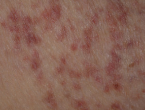
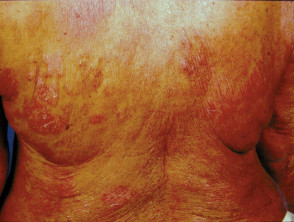
Cutaneous T-cell lymphoma
Cutaneous T-cell lymphoma (CTCL) is a lymphoproliferative disorder that manifests mainly in the skin. The most common form of cutaneous lymphoma is mycosis fungoides.
Skin manifestations
- Mycosis fungoides: single or multiple scaly patches, plaques and tumours. Varying in size. Lesions can be orange to dusky violet-red or hypopigmented
- Sézary syndrome: erythroderma/ exfoliative dermatitis
Associated features include severe pruritus, hair loss, palmoplantar keratoderma (thickening of the skin of palms of hands/soles of feet) and secondary bacterial infection.
Cutaneous T-cell lymphoma
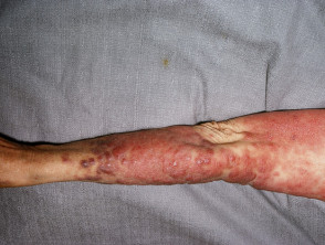
Cutaneous B-cell lymphoma
B-cell lymphomas can also present in the skin without evidence of spread elsewhere at the time they are diagnosed. These are known as primary cutaneous B cell lymphomas (PCBCL), including primary cutaneous marginal-zone lymphoma. Some of these are Epstein–Barr virus-associated lymphoproliferative disorders.
One particularly aggressive form of PCBCL is primary cutaneous large B cell lymphoma, leg type. Patients tend to be elderly and present with solitary or clustered, ulcerated, bluish, erythematous plaques or tumours located on the legs. This may be misdiagnosed as leg ulcers related to chronic venous insufficiency.
Cutaneous B-cell lymphoma
Plasma cell and immunoglobulin disorders
Multiple myeloma can rarely present with cutaneous plasmacytoma.
POEMS syndrome presents with Polyneuropathy, Organomegaly, Endocrinopathy, M protein and Skin changes. There is plasma cell proliferation and signs are mediated by overproduction of cytokines and other inflammatory markers.
Cryoglobulins are immunoglobulins that precipitate in cold temperatures. They result in palpable purpura and hyperviscosity syndrome with Raynaud phenomenon. Cryoglobulinaemia is classified as:
- Type 1 (due to underlying B-cell malignancy)
- Mixed (Type 1 + Type II) associated with B-cell malignancy or autoimmune diseases.
Cold agglutins are cold-sensitive immunoglobulins directed against antigens on red cells. They can lead to Raynaud phenomenon and acrocyanosis.
Skin signs of plasma cell disease
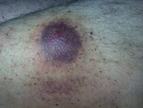
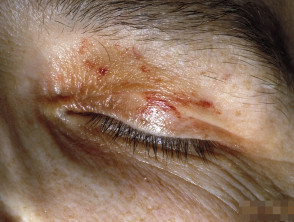
Amyloidosis
Amyloidosis refers to a group of protein-folding disorders where abnormal proteins are deposited in various tissues and organs, including the skin. The commonest of these is AL amyloidosis (light chain).
Skin manifestations
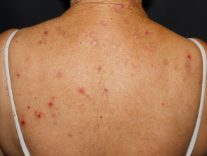
- Petechiae (pinpoint red blood spots)
- Pinch purpura (blood spots as a result of stroking skin)
- Ecchymoses (flat bluish/purplish blood spots, larger than petechiae)
- Waxy induration of the skin
- Macroglossia (enlarged tongue)
Skin signs of amyloidosis
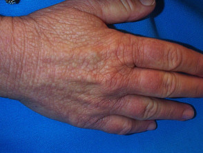
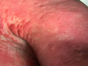
Haematopoietic stem cell transplantation
Stem cells for transplantation can be sourced from the blood, bone marrow or umbilical cord blood. Haematopoietic stem cell transplantation is an option to treat haematological malignancies where other treatment options have failed, for instance, diffuse large B cell lymphoma and myeloproliferative disorders.
Complications from haematopoietic stem cell transplantation can arise from side effects of immunosuppression, or from the person’s immune system attacking the ‘foreign’ donated cells (graft versus host disease, GVHD). Any organ in the body can be affected by acute or chronic GVHD (eg liver, lung, joints); however, the skin is most commonly affected.
Skin manifestations
- Erythema of palms, soles and ears
- Bullae
- Eosinophilic folliculitis
- Poikiloderma (thinning of the skin with areas of increased and loss of skin pigment) – chronic GVHD
Extracorporeal photopheresis (ECP) is used to treat cutaneous GVHD in stem cell transplant patients.
Coagulation disorders
Disorders of coagulation result in either excessive blood clotting or excessive bleeding.
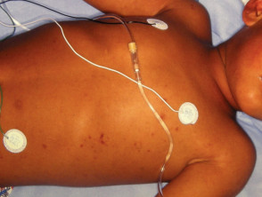
Disseminated intravascular coagulation (DIC) results in excessive clotting. It is triggered by severe infection, such as meningococcal disease, or illness, such as disseminated cancer. Severe DIC can consume all the clotting factors, which conversely leads to bleeding.
Skin manifestations are:
- Petechiae
- Ecchymoses
- Bleeding or oozing from wound sites, intravenous lines or catheters.
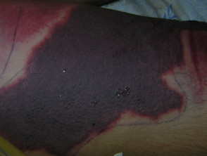
Thrombocytopenia leads to a bleeding tendency. For example in immune thrombocytopenia, platelet levels are reduced to a varying degree by circulating antibodies against them. Thrombocytopenia can also be due to bone marrow disease, Wiskott Aldrich syndrome and drugs.
Skin manifestations are:
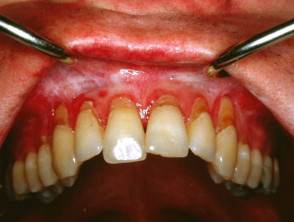
- Oral or mucocutaneous petechiae
- Ecchymoses
- Pigmented purpura.
Other coagulation disorders with cutaneous signs include:
Skin signs of coagulopathy
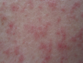
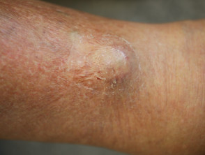
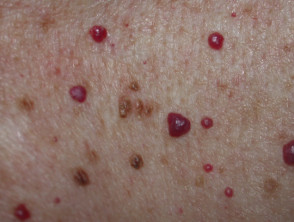
Inflammatory disorders associated with haematological disease
Skin manifestations are:
- Generalised hyperpigmentation
- Haemangiomas (tumour of blood vessels)
- Hypertrichosis (excessive hair growth)
- Telangiectasia (spider-like reddish/purplish capillaries)
- Cutaneous vasculitis.
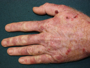
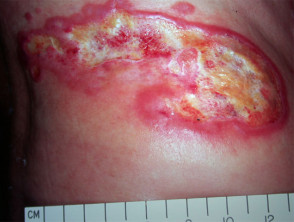
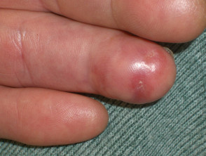
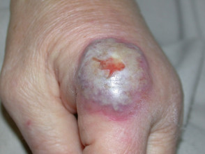
Sweet syndrome (acute febrile neutrophilic dermatosis) is an acute inflammatory skin eruption that is accompanied by fevers and leukocytosis. It can be associated with infections, inflammatory bowel disease, haematological cancers and certain drugs (eg antibiotics). A localised variant of Sweet syndrome is known as neutrophilic dermatosis of the dorsal hands.
Skin manifestations
- Tender reddish/purplish papules or nodules – can be single or multiple. Lesions may appear bullous, ulcerated or mimic pyoderma gangrenosum
- Pustules (pus-filled blisters)
- Tender, erythematous nodules on extremities (can mimic erythema nodosum)
- Cutaneous pathergy (skin lesions appearing at sites of trauma)
Inflammatory skin disease associated with haematological disease
Adverse reactions to drugs
Drugs used to treat specific haematological conditions can have toxicities that manifest in the skin. Examples include:
- Porphyria cutanea tarda and vitiligo due to interferon alpha in the treatment of chronic myeloid leukaemia (CML)
- Psoriasiform dermatitis, pruritus, alopecia and photosensitivity due to tyrosine kinase inhibitors like imatinib used to treat CML, cutaneous mastocytosis and sclerodermic GVHD
- Hydroxyurea can lead to dry skin, actinic keratoses, squamous cell carcinoma, leg ulcers, hyperpigmentation, and nail changes.
Adverse reactions to drugs used in haematology