What is cutaneous squamous cell carcinoma?
Cutaneous squamous cell carcinoma is a common type of keratinocytic or nonmelanoma skin cancer. It is commonly found on sun-exposed areas of skin. It can be invasive and metastasise. It is also known as cutaneous squamous cell carcinoma, and commonly abbreviated to SCC.
What are the clinical features of squamous cell carcinoma?
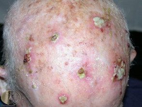
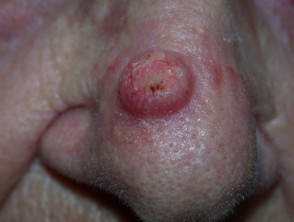
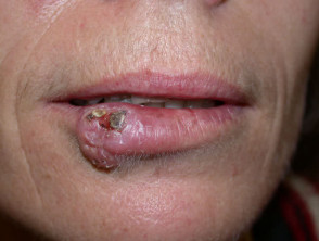
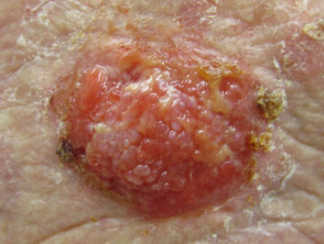
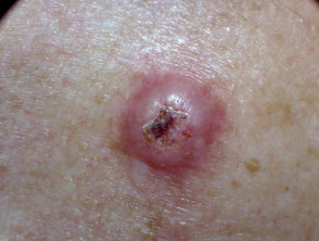
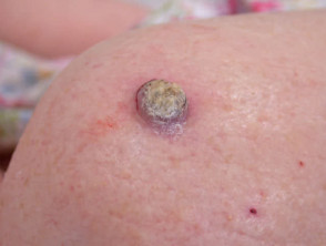
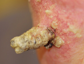
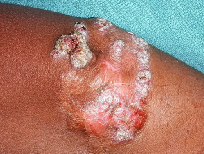
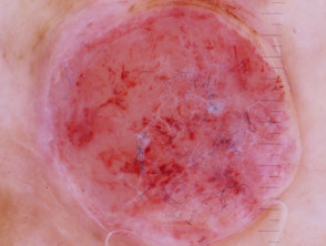
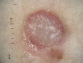
Cutaneous squamous cell carcinoma presents clinically as an enlarging, irregular, keratinous nodule or a firm erythematous plaque that frequently ulcerates.
They usually arise within pre-existing actinic keratosis or intraepidermal carcinoma.
- They grow over weeks to months
- They may ulcerate
- They are often tender or painful
- Located on sun-exposed sites, particularly the face, lips, ears, hands, forearms, and lower legs
- Size varies from a few millimetres to several centimetres in diameter
- They are rarely pigmented.
What are the dermoscopic features of squamous cell carcinoma?
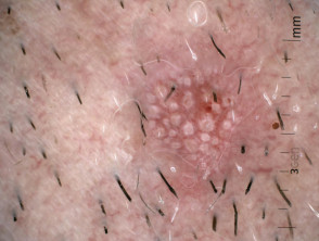
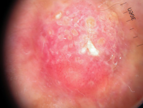
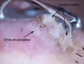
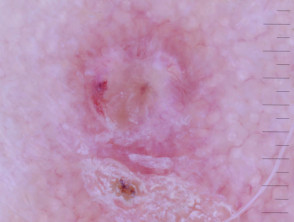
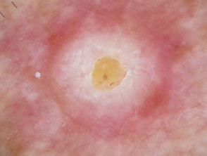
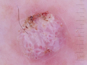
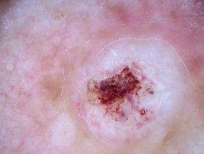
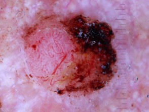
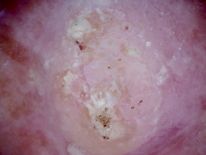
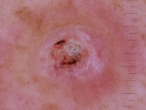
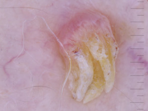
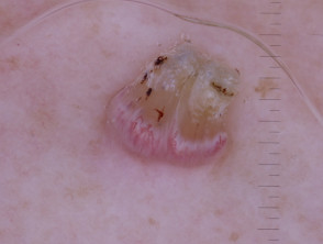
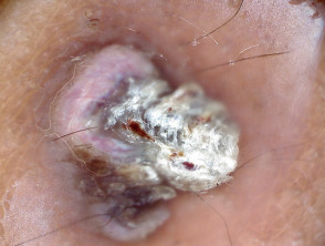
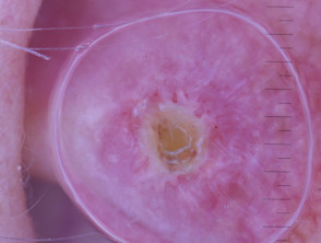
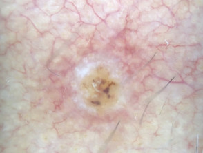
The common dermoscopic features of cutaneous squamous cell carcinoma are:
- White circles
- White structureless areas
- Looped vessels
- Central keratin
- Pink or red background in poorly differentiated or rapidly growing tumours.
White structureless areas and white circles in squamous cell carcinoma dermoscopy White circles in squamous cell carcinoma dermoscopy White circles and surface keratin seen in squamous cell carcinoma dermoscopy White structures and surface keratin in squamous cell carcinoma dermoscopy Loop vessels seen in squamous cell carcinoma dermoscopy White structureless areas and central keratin in squamous cell carcinoma dermoscopy White structureless areas and surface keratin in squamous cell carcinoma dermoscopy White structureless areas, central keratin and loop vessels in squamous cell carcinoma dermoscopy Poorly differentiated squamous cell carcinoma dermoscopy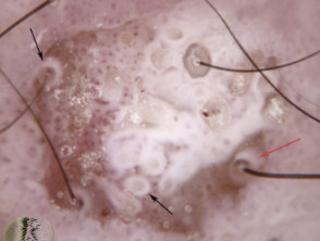
Pigmented cutaneous squamous cell carcinoma has brown, blue or grey areas.
What is the dermoscopic differential diagnosis of squamous cell carcinoma?
The dermoscopic differential diagnosis of cutaneous squamous cell carcinoma includes its variants:
- Intraepidermal carcinoma (also known as IEC, squamous cell carcinoma in-situ and Bowen's disease)
- Cutaneous horn
- Actinic keratosis
- Keratoacanthoma.
Other conditions to consider include:
- Viral wart
- Seborrhoeic keratosis
- Basal cell carcinoma (especially if pigmented)
- Melanoma
- Blastomycosis
- Chondrodermatitis nodularis helicis.
Intraepidermal carcinoma dermoscopy Macro image of a cutaneous horn Actinic keratosis dermoscopy Keratoacanthoma dermoscopy Keratoacanthoma dermoscopy Verruca vulgaris dermoscopy Verruca vulgaris dermoscopy Macro image of chromoblastomycosis Chondrodermatitis nodularis helicis dermoscopy Basal cell carcinoma dermoscopy Amelanotic melanoma, Breslow 3.2mm, CL IV dermoscopy Amelanotic melanoma dermoscopy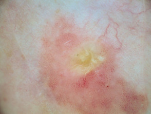
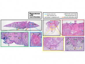
What is the histological explanation for squamous cell carcinoma?
Histologically, there is a proliferation of atypical keratinocytes within the dermis.