What is staphylococcal scalded skin syndrome?
Staphylococcal scalded skin syndrome (SSSS) is a rare, severe, superficial blistering skin disorder which is characterised by the detachment of the outermost skin layer (epidermis). This is triggered by exotoxin release from specific strains of Staphylococcus aureus bacteria.
The blistering of large areas of skin gives the appearance of a burn or scalding, hence the name staphylococcal scalded skin syndrome. SSSS is often used interchangeably with the eponymous name Ritter von Ritterschein disease (Ritter disease), particularly when it presents in newborn children.
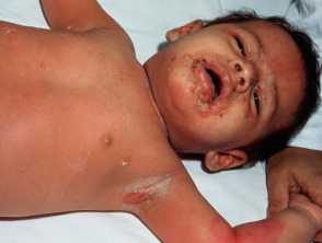
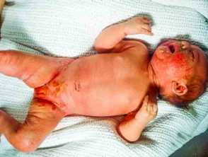
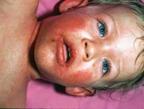
Superficial blistering over the axillae, face and chest due to staphylococcal scalded skin syndrome Superficial erosions in the napkin area and widespread erythema in staphylococcal scalded skin syndrome
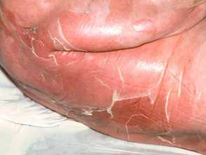
Erythema and wrinkling of the necrotic epidermis in staphylococcal scalded skin syndrome
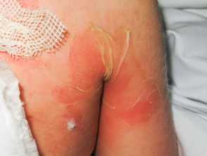
A post-operative Staphylococcal wound infection was followed by this eruption - widespread partial thickess epidermal necrosis with normal mucous membranes due to staphylococcal scalded skin syndrome
Staphylococcal scalded skin syndrome Erythema, scaling and crusting in an infant with staphylococcal scalded skin syndromeStaphylococcal scalded skin syndrome
Who gets staphylococcal scalded skin syndrome?
SSSS is predominantly seen in children younger than 5 years with a peak age being reported between 2 to 3 years of age. The higher incidence in this age group is thought to be a consequence of an immature:
- Immune system: lack of protective antibodies to exotoxins
- Renal clearance system: reduced toxin clearance.
Although rare, cases have been reported in older children and adults, and would characteristically involve those who are immunosuppressed or have severe renal impairment/chronic renal disease.
The estimated prevalence of SSSS based on studies undertaken in Europe has been reported to range from 0.09 to 0.56 cases per million people, with an equal male to female ratio seen in children.
What causes staphylococcal scalded skin syndrome?
SSSS starts from a localised infection caused by toxigenic Staphylococcus aureus (approximately 5% of strains). The following then occurs:
- S. aureus releases two exotoxins (exfoliative toxins A and B)
- These toxins bind to a specific desmosome present in the epidermis known as desmoglein-1
- Desmoglein-1 is responsible for maintaining cell-to-cell adherence within the epidermis and preserving skin integrity
- The desmoglein-1 protein is broken down leading to the skin cells becoming loose and “unstuck”
- Loss of the epidermal layer and associated blistering occurs.
Depending on disease severity, the resultant effect can range from a localised area of skin loss to total body blistering and sloughing. Desmoglein-1 is absent from mucosal epithelium, which explains why mucosae are unaffected in SSSS.
The initial localised infection often starts from sites such as the ears, eyes (conjunctiva) or throat, especially in children, or infected wounds. These toxins then spread to the skin via the circulating blood and target the desmoglein-1 protein in the epidermis leading to the blistering and sloughing typified by the condition.
What are the clinical features of staphylococcal scalded skin syndrome?
SSSS tends to start with nonspecific symptoms in children; this may include irritability, lethargy, and fever. Within 24–48 hours, a painful widespread red rash develops on the skin followed by the formation of large, fragile, fluid-filled blisters (bullae). These can rupture easily leaving tender patches of skin that look like a burn.
Typical SSSS cutaneous features include:
- A red rash with wrinkled tissue or paper-like consistency
- Typically starts on the face and flexural regions (groin, axillae, and neck), then spreads rapidly to other parts of the body including the arms, legs, and trunk.
- In newborns, lesions can be found around the umbilical cord.
- Following the rash, the formation of large fluid-filled blisters
- Frequently occur in areas of friction (such as axillae, groin, and buttocks), the centre of the face and body orifices (such as the nose and ears).
- Fluid contents range from a sterile cloudy fluid to frank yellow pus.
- Blisters easily rupture leading to the top layer of the skin (epidermis) peeling off easily, often in large sheets.
- Exposure of the underlying, moist, reddish tissue leaves the skin with a burned-like appearance.
- Gentle rubbing of the skin causes exfoliation (Nikolsky sign is positive).
There is typically no mucous membrane involvement in SSSS, which helps to distinguish it from toxic epidermal necrolysis (TEN), where mucosal changes are usual.
How do clinical features vary in differing types of skin?
Erythema in the initial phase of SSSS is less easily appreciated in skin or colour.
What are the complications of staphylococcal scalded skin syndrome?
Despite the alarming appearance, children who contract SSSS generally experience complete recovery within two weeks if diagnosed and treated promptly. However, if left untreated or treatment is unsuccessful, a number of complications can occur. These are typically due to the loss of the protective skin barrier, which include:
- Scarring
- Loss of bodily fluids and salts leading to dehydration and electrolyte imbalance
- Hypothermia
- Secondary infections such as sepsis, cellulitis, and pneumonia
- Renal failure.
The mortality rate from SSSS in children is extremely low (1–5%), unless they develop secondary sepsis or have an underlying serious medical condition that complicates the disease course. The mortality rate in adults is higher (50–60%), likely due to an underlying condition which increases their predisposition to complications in itself.
How is staphylococcal scalded skin syndrome diagnosed?
A history and physical examination are often sufficient for diagnosis. Tests that may be used include:
- Skin swabs can be taken from the suspected source of infection and/or the blister fluid to confirm the presence of S. aureus. However, these can frequently be negative given the condition is toxin mediated.
- Blood cultures are undertaken when sepsis is of concern.
- Tzanck smear
- Skin biopsy is occasionally undertaken to exclude other causes of blisters, and occasionally is a justification for frozen section histology
- SSSS reveals non-inflammatory intra-epidermal splitting at the granular layer.
What is the differential diagnosis for staphylococcal scalded skin syndrome?
- Steven–Johnsons syndrome/Toxic Epidermal Necrolysis (SJS/TEN)
- Bullous impetigo
- Drug hypersensitivity reaction
- Viral exanthem
- Thermal burns.
What is the treatment for staphylococcal scalded skin syndrome?
SSSS is considered a dermatological emergency which requires hospitalisation and prompt treatment. This usually involves:
- Intravenous antibiotics
- First-line: a penicillinase-resistant, anti-staphylococcal antibiotic such as flucloxacillin.
- Other options include: ceftriaxone, clarithromycin (for penicillin-allergy), cefazolin, nafcillin, or oxacillin.
- Methicillin resistance (MRSA) infection: vancomycin.
If there is a good response, oral antibiotics can be substituted within several days. The patient may then be discharged from hospital to continue treatment at home.
Supportive treatments for SSSS include:
- Pain relief
- Paracetamol with addition of alternative analgesia such as ibuprofen or oral morphine if required whilst the skin heals.
- Monitoring and maintaining fluid and electrolyte intake:
- Intravenous fluids should be considered in young children and for those with widespread skin involvement.
- Skincare
- Gentle washing of the skin at least once a day with a soap substitute
- Application of greasy emollients such as 50:50 white soft paraffin/liquid paraffin or petroleum jelly to soothe the skin and help with healing.
- Burns dressings may be required in some areas.
Despite the initial alarming appearance of SSSS, children often make an excellent recovery with complete healing usually occurring within 5–7 days of starting treatment.
How do you prevent staphylococcal scalded skin syndrome?
If there is an outbreak of SSSS in either a neonatal care unit or childcare facility, the possibility of a staphylococcal carrier in the vicinity should be investigated. Identification of the healthcare worker, childcare worker, parent or visitor colonised or infected with Staphylococcus aureus is key to managing the problem. Once identified these individuals should be treated with oral antibiotics to eradicate the causative organism. To prevent further infections these places should employ strict hand washing with antibacterial soap or sanitisers.
What is the outcome of staphylococcal scalded skin syndrome?
Despite the alarming appearance, children who contract SSSS generally experience complete recovery within two weeks if diagnosed and treated promptly. However, if left untreated or treatment is unsuccessful, a number of complications can occur (see above).
Mortality is low in children (1–5%) but much higher in adults (50–60%); due to underlying comorbidities.