Introduction
Tinea nigra is a superficial mycosis caused by Hortaea werneckii.
Histology of tinea nigra
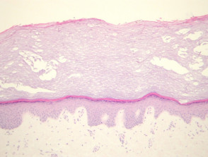
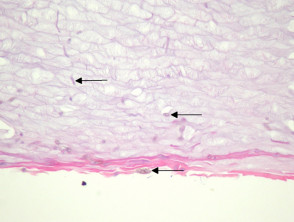
The epidermis and dermis of the acral skin infected with tinea nigra are largely unremakarkable at low power magnification (figure 1). In the superficial aspects of the stratum corneum, numerous short, segmented hyphae and spores are seen These organisms have a characteristic brown-yellow colour on routine haematoxylin and eosin sections (figure 2, arrows).
Tinea nigra pathology
Special studies for tinea nigra
The organisms are typically easy to identify at high power with haematoxylin and eosin sections. PAS and GMS stains can also be used to highlight the morphology.
Differential diagnosis of tinea nigra pathology
Tinea – The fungal forms of tinea (dermatophyte infection) lack the characteristic brown-yellow pigment and typically shows more of an inflammatory response.