What is trichilemmoma?
Trichilemmoma, also called tricholemmoma is a benign tumour originating from the outer root sheath of the hair follicle. Diagnosis depends on careful histopathological examination of a skin biopsy.
Trichilemmoma often occurs alongside other skin lesions such as trichoblastoma, sebaceous adenoma and sebaceous naevus. Occasionally trichilemmoma may appear as a new growth within a sebaceous naevus lesion.
Multiple trichilemmoma on the face is often associated with Cowden disease, a rare inherited condition characterized by multiple types of skin tumours which can be found throughout different body systems. Although multiple trichilemmoma associated with Cowden disease is very rare, it is important to re-evaluate patients for this disease if a diagnosis of trichilemmoma is confirmed.
The underlying cause of trichilemmoma is unknown but because of its similarity to a viral wart/verruca, it has been postulated that it may come from a viral infection such as the human papillomavirus (HPV). To date there is no evidence for this.
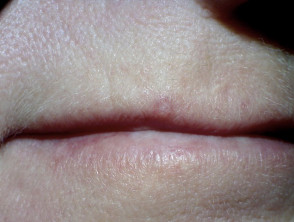
What are the clinical features of trichilemmoma?
Trichilemmoma typically presents as a solitary papule or mass of small skin-coloured papules that are 1-5 cm in diameter. These lesions slowly grow over time and tend to form small plaques that may resemble a wart/verruca or a cutaneous horn. They most commonly occur around the central part of the face, ears and neck, but also occur on the forearms and hands.
Solitary trichilemmomas are relatively common benign follicular tumours and occur in both male and females usually between 20-80 years of age. Desmoplastic trichilemmomas, a subtype of trichilemmoma, mainly occurs in white males around 50 years old. Lesions of this subtype are usually less than 1 cm in diameter and occur mainly on the face, neck and scalp, and sometimes on the chest and vulva.
Trichilemmoma
How is trichilemmoma diagnosed?
A small biopsy (when a tiny piece of skin is removed under local anaesthetic) is the only definitive diagnosis for trichilemmoma. The histology of trichilemmoma will differentiate it from other skin tumours that have similar clinical presentations, such as trichoepithelioma, trichofolliculoma and basal cell carcinoma.
Where there are multiple lesions on the face, once a diagnosis of trichilemmoma is confirmed, patients should be completely examined for evidence of Cowden disease.
What is the treatment of trichilemmoma?
Trichilemmoma is a benign follicular tumour that requires no treatment. Occasionally lesion(s) may be removed for cosmetic reasons or if they occur in functionally sensitive areas. The main reason for surgery is to establish the correct diagnosis and to ensure that a potential malignancy, such as trichilemmal carcinoma, is not left untreated.
Treatment options include.