What is venous insufficiency?
Venous insufficiency occurs when the normal flow of blood from the superficial veins to the heart via the perforating deep veins in the lower limbs is impaired, resulting in chronic venous congestion. It can be classified as superficial vein insufficiency, perforating, or deep vein insufficiency.
Venous insufficiency
Who gets venous insufficiency?
Venous insufficiency is common, affecting all races and both sexes. Estimates suggest rates as high as 50% in some populations. A US study found ethnic whites had a higher rate of venous insufficiency compared to Hispanics, African Americans, and Asians. The Edinburgh Vein Study reported the incidence of chronic venous insufficiency was similar in men and women. Prevalence increases with age, obesity, a family history of varicose veins, and multiple pregnancies.
What causes venous insufficiency?
The venous system in the lower legs consists of a low-pressure superficial network connected by perforating veins to a high-pressure deep network. Venous blood flows from the superficial to deep veins by the action of the calf muscle pump. Retrograde flow is prevented by competent valves.
Chronic venous insufficiency can be caused by:
- Venous factors
- Incompetent valve in the superficial, perforating, and deep systems
- External compression of veins
- Deep venous obstruction
- Previous deep vein thrombosis
- Impaired calf muscle pump
- Immobility including lack of physical exercise and paralysis
- Joint disease particularly of the ankle
- Obesity
- Prolonged standing or sitting
- Previous leg injury.
Once venous hypertension is established, the cutaneous features of chronic venous insufficiency are caused by:
- Leukocytes trapped in capillaries release proteolytic enzymes and oxygen free radicals which damage the capillary basement membrane
- Plasma proteins, such as fibrinogen, leak into the surrounding tissues, forming a fibrin cuff which reduces oxygen delivery to the tissues
- Inflammation and tissue loss.
What are the clinical features of venous insufficiency?
Superficial venous insufficiency can be asymptomatic but may cause aching, cramping, throbbing, burning, or heaviness in the leg. Pain is typical of deep venous insufficiency. Symptoms improve with leg elevation.
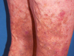
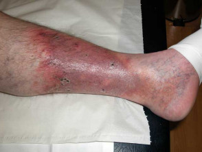
Clinical signs of chronic venous insufficiency include:
- Pitting oedema around the ankle, worse at the end of the day
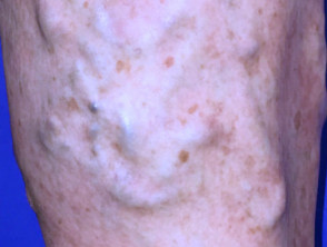
- Corona phlebectatica paraplantaris (malleolar flare) on the inner aspect of the ankle and instep – fan-shaped distribution of blue and red telangiectases (intradermal and superficial venules), capillary stasis spots (red-purple coin-shaped areas formed by papillary capillaries), and venous cups (dilated converging veins on the instep)
- Telangiectasia adjacent to perforating veins
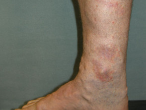
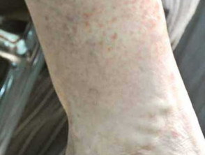
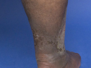
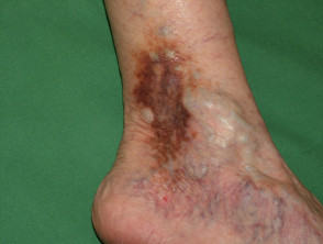
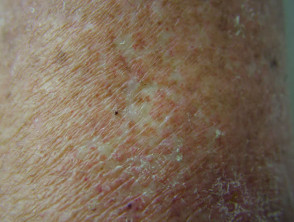
- Pigmentation can be pinpoint or a patchy rusty-red due to extravasation of red blood cells and haemosiderin accumulation in the dermis
- Varicosities.
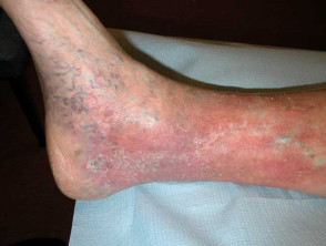
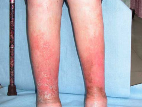
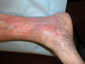
Clinical features of venous insufficiency
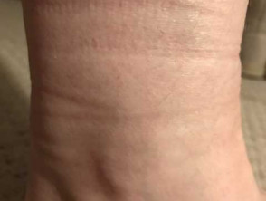
Pigment changes in venous insufficiency
How do clinical features vary in differing types of skin?
Venous insufficiency in Thai patients has been reported to be less likely to be associated with pain, oedema, inflammation, and visible varicose veins compared to ethnic whites.
What are the complications of venous insufficiency?
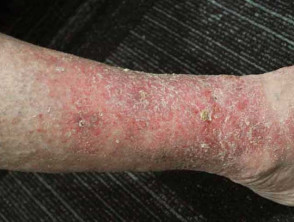
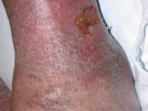
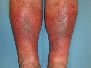
Venous eczema
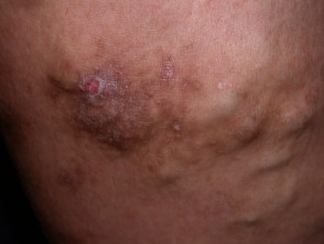
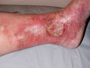
Stasis ulcers: active and healed
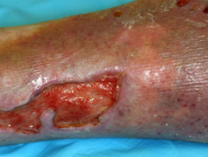
Complications of venous insufficiency
How is venous insufficiency diagnosed?
Venous insufficiency is usually diagnosed on history and examination. Varicose veins may require the patient to be standing to appreciate. Duplex ultrasonography is the preferred investigation to demonstrate reflux and communications between the deep and superficial venous networks.
The CEAP (Clinical, Etiologic, Anatomic, Pathologic) system is used categorise and classify venous insufficiency but is unhelpful as a severity score. [see details Varicose veins]
The corona phlebectatica paraplantaris is a clinical sign of severe venous stasis. Specifically, blue telangiectases and capillary stasis spots may be the most useful correlates with the severity of chronic venous insufficiency.
What is the differential diagnosis for venous insufficiency?
- Oedema — lipoedema, lymphoedema
- Pigmentation — capillaritis
- Dermatitis — contact dermatitis, asteatotic eczema, discoid eczema
What is the treatment for venous insufficiency?
General measures
- Soap and detergent avoidance
- Regular use of moisturisers for dryness and itch
- Lifestyle changes — regular exercise, leg elevation, weight loss
Specific measures
- Graduated compression hosiery or bandages [see Compression therapy]
- Telangiectasia — laser therapy, sclerotherapy
- Varicose veins — sclerotherapy, endovenous laser therapy, radiofrequency ablation, surgery [see Leg vein therapies]
What is the outcome for venous insufficiency?
Venous insufficiency is inexorably progressive. The rate of complication development is not known.
Most patients with chronic venous insufficiency will experience at least intermittent episodes of venous eczema.
Leg ulceration is the most important complication and is a major burden for health systems throughout the world. Estimates suggest 4% of patients with varicose veins will develop venous leg ulcers in their lifetime.