What is idiopathic guttate hypomelanosis?
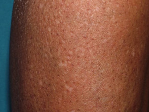
Idiopathic guttate hypomelanosis is a common acquired form of leukoderma that presents as small flat pale or white spots on the sun-exposed limbs.
Idiopathic guttate hypomelanosis


Who gets idiopathic guttate hypomelanosis?
Idiopathic guttate hypomelanosis can affect both sexes, all races, and all skin phototypes. Women and patients with skin of colour are most likely to present for medical attention.
Idiopathic guttate hypomelanosis becomes more common with age, affecting <50% in the fourth decade (31–40 years), 50–80% of people over 40 years of age, and >90% in the ninth decade (81–90 years). It has been reported uncommonly in children and teens. Familial cases are common.
What causes idiopathic guttate hypomelanosis?
The aetiopathogenesis of idiopathic guttate hypomelanosis is probably multifactorial:
- Skin ageing
- Chronic sun exposure
- Genetic factors
- Other — trauma, autoimmune factors.
Abnormal keratinocyte phagocytosis has been demonstrated resulting in reduced transfer of melanin from melanocytes.
What are the clinical features of idiopathic guttate hypomelanosis?
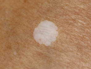
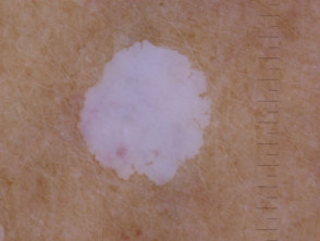
Idiopathic guttate hypomelanosis typically presents as smooth pale (hypopigmented) or white (depigmented) macules 2–5mm (range up to 1.5cm) in diameter most commonly on the sun-exposed aspects of the forearms, shins, and V of chest. Lesions are usually multiple and asymptomatic.
Idiopathic guttate hypomelanosis


Three morphological variants have been described:
- Solitary or multiple hypopigmented macules on a background of sun-damaged skin
- Solitary white stellate sclerotic macule
- Small hypopigmented macules with a scalloped margin and hyperkeratotic surface.
Wood lamp examination emphasises the loss of pigment.
How do clinical features vary in differing types of skin?
Idiopathic guttate hypomelanosis is more obvious in skin of colour.
Dermoscopy of idiopathic guttate hypomelanosis
- Well-defined white structureless area with ‘glow’
- Absent pigment network
- Various patterns and shapes described
[for a more detailed description and images see Idiopathic guttate hypomelanosis dermoscopy]
Macro and dermoscopy images of idiopathic guttate hypomelanosis

What are the complications of idiopathic guttate hypomelanosis?
Idiopathic guttate hypomelanosis is a benign condition of cosmetic significance. It can impact quality of life particularly in skin of colour.
How is idiopathic guttate hypomelanosis diagnosed?
Idiopathic guttate hypomelanosis is usually a clinical diagnosis.
Skin biopsy may be performed to rule out other conditions and histology is specific:
- Flat thin epidermis with basket-weave hyperkeratosis and loss of rete
- Absent or decreased melanin in epidermis
- Normal or reduced melanocyte number and activity; large melanocytes with retained melanin and small retracted dendritic processes
- Small foci of retained melanin in the basal layer; ‘skip lesions’.
What is the differential diagnosis for idiopathic guttate hypomelanosis?
- Vitiligo
- Lichen sclerosus and guttate morphoea
- Post-inflammatory hypopigmentation such as following cryotherapy
- Contact leukoderma
What is the treatment for idiopathic guttate hypomelanosis?
Idiopathic guttate hypomelanosis usually does not require treatment apart from reassurance as to the benign nature of the condition.
Sun protection is recommended.
Treatment options:
- Cryotherapy — 5 second, single session, repigmentation visible by 4 months
- Topical treatments — topical steroid, tacrolimus, topical retinoids
- Procedural treatments — chemical peel, excimer laser, skin grafting.
What is the outcome for idiopathic guttate hypomelanosis?
Idiopathic guttate hypomelanosis does not spontaneously repigment. With age, lesions may slowly enlarge and increase in number.