What is malassezia?
Malassezia refers to a group of basidiomycetous (club-shaped) yeasts of the genus Malassezia, which has several different species. Species names include:
- M. dermatis
- M. equi
- M. furfur
- M. globosa
- M. obtusa
- M. pachydermatis
- M. restricta
- M. slooffiae
- M. sympodialis
- M. ovalis (also known as Pityrosporum ovale)
What skin conditions are associated with malassezia?
Skin conditions caused or aggravated by infection by malassezia include:
- Pityriasis versicolor – most often due to M. globosa, M. sympodialis and M. furfur
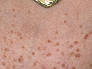
- Malassezia folliculitis due to the yeast growing in the hair follicles where they produce inflammation
- Steroid acne
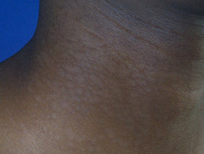
- Seborrhoeic dermatitis, dandruff, sebopsoriasis and facial or scalp psoriasis - – most often due to M. restricta and M. globosa.
- Neonatal cephalic pustulosis, a pustular eruption on young babies that resembles infantile acne
- Pseudochromhidrosis
- Possibly, some cases of confluent and reticulated papillomatosis, a pigmented eruption occurring mainly on the chest, back and neck of adolescent girls
- Some facial atopic dermatitis; in this case there may be specific IgE antibodies directed against Malassezia and positive prick tests to the organism
- Rarely, invasive pityrosporosis in immunodeficient individuals.
Malassezia infections

How are skin conditions associated with malassezia diagnosed?
The diagnosis of skin conditions associated with malassezia is often made clinically but can be confirmed when skin scrapings reveal malassezia (see laboratory tests for fungal infection). Microscopy of malassezia, using potassium hydroxide (KOH) preparations, shows clusters of yeast cells and long hyphae. The appearance is said to be like 'spaghetti and meatballs'. The hyphae filaments used to be called 'Malassezia' and the yeast forms were called 'Pityrosporum', but mycologists eventually realised they were the same bimorphic organism.
Malassezia species are difficult to grow in the laboratory so scrapings may be reported as 'culture-negative'. The yeast grows best if a lipid such as olive oil is added to Littman agar culture medium.
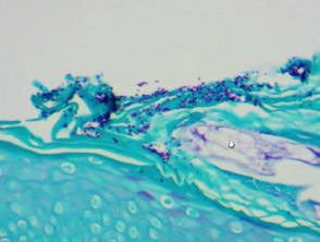
Yeasts may also be detected within the stratum corneum on histopathology of a skin biopsy; they are best seen using special stains such as periodic acid-Schiff (PAS).
Malassezia yeast cells in stratum corneum
What are predisposing factors to malassezia proliferation?
Malassezia species inhabit the skin of about 90% of adults without causing harm. In some people, the yeast suppresses the body's expected immune response to it allowing it to proliferate and cause a skin disorder, often with very little inflammatory response. When malassezia is associated with dermatitis, it is thought that irritating metabolites of the yeasts may be responsible (free fatty acids are hydrolysed from triglycerides).
Predisposing factors to Malassezia skin disease include:
- Humidity
- Sweating – hence pityriasis versicolor is common in tropical areas
- Oily skin (seborrhoea) – hence it is found mainly on scalp, face and upper trunk
- Acne and its treatment with oral antibiotics such as tetracyclines
- Immunodeficiency (eg, HIV infection), systemic corticosteroids, or immunosuppression by medications.
The yeasts produce chemicals that reduce the pigment in the skin, causing whitish patches. These include azelaic acid, pityriacitrin and malassezin. Azelaic acid is a useful treatment for some skin disorders such as acne and rosacea.
Malassezia may fluoresce on exposure to ultraviolet light (Wood lamp). This is due to another chemical, pityrialactone.
What is the treatment of skin conditions associated with malassezia?
Consult DermNet's pages on the individual skin conditions to learn about treatment.
In general, malassezia infections are treated with topical or oral antifungal agents, such as ketoconazole shampoo and oral fluconazole. Seborrhoeic dermatitis may also be treated with topical steroids.