What is atrophie blanche?
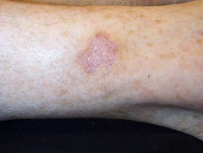
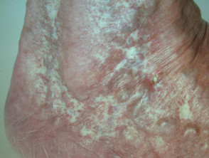
Atrophie blanche (white atrophy) is the name given to a particular type of angular scar arising on the lower leg or foot. It occurs after a skin injury, when the blood supply is poor and healing is delayed.
Atrophie blanche
What causes atrophie blanche?
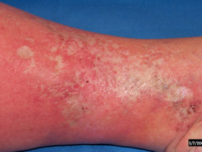
Atrophie blanche most often occurs in middle-aged women. It is most often associated with venous insufficiency. It is the hallmark of livedoid vasculopathy, and may also follow ulceration due to:
- Cutaneous small vessel vasculitis
- Diabetic vascular disease
- Any wound on the lower leg, such as following cryotherapy or curettage and cautery in the treatment of skin cancer.
Atrophie blanche is due to occlusion of small blood vessels in the middle and deep dermis, which prevents normal healing. Blood vessel occlusion may be due to:
- Microthrombi
- Defective endothelium
- Enhanced production of fibrin
- Fibrin cuff formation
- Increased platelet aggregation
- White-cell trapping
- Autoantibodies
- Primary vasculitis
- Infection.
What are the clinical features of atrophie blanche?
Atrophie blanche is characterised by:
- Star-shaped or polyangular, ivory-white depressed atrophic plaques
- Prominent red dots within the scar due to enlarged capillary blood vessels
- Surrounding hyperpigmentation due to haemosiderin deposition.
What is the treatment of atrophie blanche?
Treatment is directed to the underlying disease process that leads to atrophie blanche. For example, in livedoid vasculopathy, drugs are used that halt platelet aggregation and stimulate fibrinolysis.
Compression therapy may speed up healing of wounds on the lower leg, particularly in venous disease, and thus reduce the severity of atrophie blanche scar formation.