Introduction
Demographics
Causes
Clinical features
Complications
Diagnosis
Differential diagnoses
Treatment
Outcome
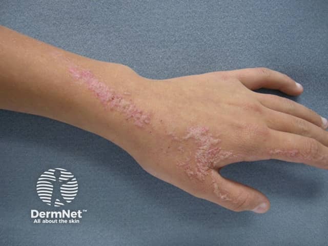
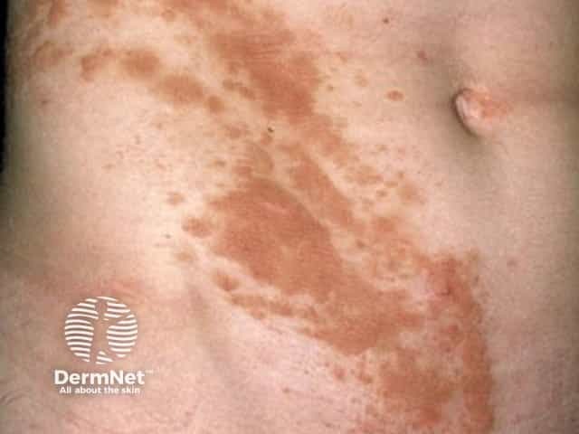
Inflammatory linear verrucous epidermal naevus (ILVEN) is a benign naevus characterised by unilateral pruritic, erythematous, and hyperkeratotic papules and plaques distributed along the Blaschko lines.
It is a rare variant of keratinocytic epidermal naevus and part of a heterogeneous group of mosaic inflammatory disorders.

ILVEN arises in the first five years of life but may be present at birth. It is more common in females with no racial predilection. Although not considered an inherited disorder, familial cases have been rarely reported.
The exact aetiology of ILVEN remains unknown, however it is understood that the abnormality resulting in ILVEN arises from a defect in the ectoderm. This is the outer layer of the embryo that gives rise to neural tissue and the epidermis. The genetic mutations that give rise to ILVEN are usually sporadic, but familial cases have been reported. ILVEN is highly heterogeneous genetically and mosaic mutations have been identified in numerous genes, including NSDHL, PMVK, HRAS, GJA1, and CARD14.
Causative genetic mechanisms include germline X-linked variants, mosaic variants due to somatic mutations, and germline mutations followed by a second-hit somatic mutation.
Genetic analysis can potentially categorise ILVEN and identify pathogenesis-directed therapies. However, many cases do not demonstrate a known genetic mutation.
Like other keratinocytic epidermal naevi, ILVEN is characterised by warty lesions that tend to group together in a linear pattern along the lines of Blaschko, unilaterally. The difference is that the lesions are erythematous, inflamed, and pruritic, sometimes intensely so. The surface of the lesions may appear eczema-like (dry, red, scratched) or psoriasis-like (red and scaly).
ILVEN most often affects one leg and may extend from the buttock to the foot. Reports have also described the involvement of the trunk, genitals, and mucosa.
The underlying ectodermal defect that results in the skin lesions seen in ILVEN may also lead to disorders of other internal organs such as the brain, eyes, and skeleton — though this is extremely rare with ILVEN.
ILVEN is diagnosed by careful history and physical examination. A skin biopsy can be helpful and may show the characteristic histological features of ILVEN including epidermal hyperplasia, papillomatosis, hyperkeratosis and parakeratosis, and diffuse or perivascular inflammatory reactions in the papillary dermis.
Genetic testing for a variety of mutations (see causes above) may help elucidate the cause and direct therapy.
While there is no cure for ILVEN, treatment aims to manage symptoms or improve appearance with varying success. These include:
ILVEN is chronic and progressive, with periods of exacerbation followed by improvement. Spontaneous regression is uncommon. Refractory cases have been reported and management can be difficult.

