Created 2008.
Dermoscopy is useful to distinguish pigmented non-melanocytic lesions from benign and malignant melanocytic lesions. There are specific features that help to distinguish these.
Careful observation has resulted in the description of the dermoscopy of many non-pigmented lesions as well, which may be sometimes helpful in diagnosis for an itchy rash.
The dermoscopic features of haemangiomas or angiomas are:
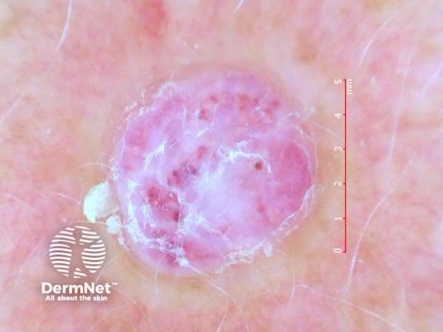
Reactive haemangioma or pyogenic granuloma has a distinct keratinised border or collarette. Vascular structures are usually present but there is no clear lacunar pattern. White linear 'rail lines' are often featured. It is not always possible to distinguish reactive haemangioma from amelanotic melanoma.
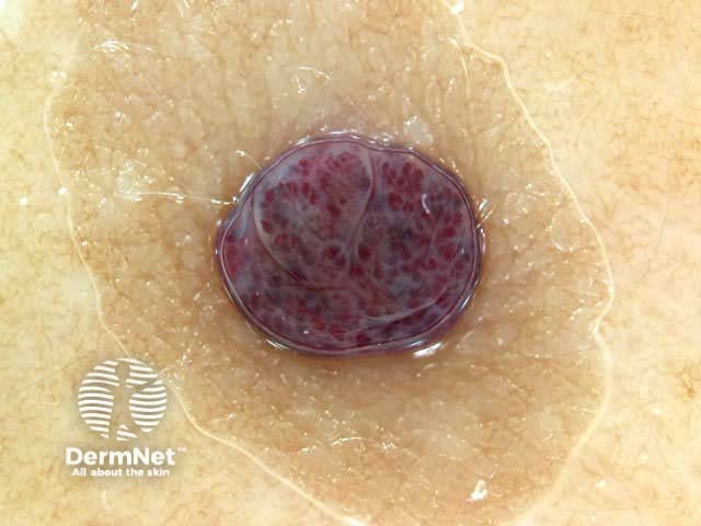
Cutaneous lymphatic malformation (formerly called lymphangioma circumscriptum) has yellowish lacunes, sometimes tinged with blood.
Kaposi sarcoma under polarised microsocopy is characterised by multicoloured rainbow pattern in association with bluish-red colour, scaling and small brown globules. The rainbow pattern is occasionally seen in melanoma and other skin lesions.


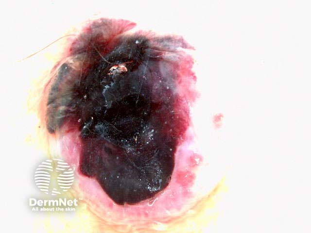
Haemorrhage can be distinguished from pigmentation due to melanin by the purple colour. On plantar surfaces (e.g. talon noir) it may appear to have a parallel ridge pattern of discolouration with peripheral reddish-black globules. It may be helpful to shave off the surface keratin – sometimes biopsy is necessary to rule out melanoma.


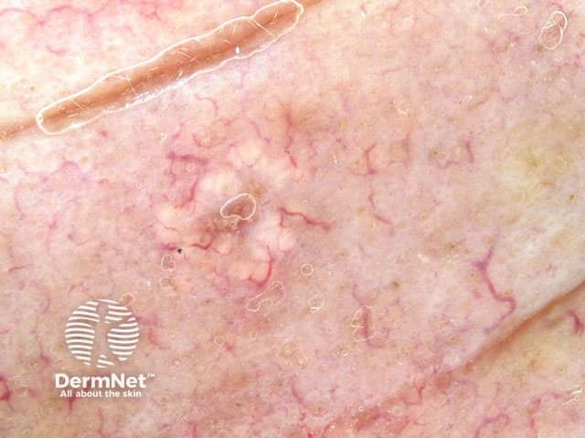
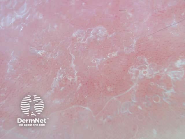
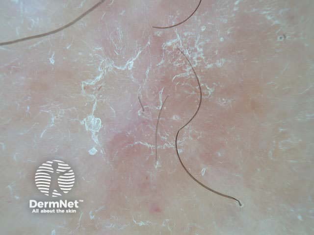
The dermatofibroma (also known as histiocytoma) is usually easy to diagnose clinically because of a firm fibrous consistency and surface dimpling on compression. Typically, dermoscopy of a dermatofibroma shows a faint network or pseudonetwork surrounding a pale amorphous area. Sometimes the central white area has white lines and brown holes (negative network). Chrystalline structures, i.e. white shiny lines, are commonly seen on polarised dermoscopy of dermatofibroma.
Haemosiderotic dermatofibroma (uncommon) is composed of numerous small vessels, extravasated erythrocytes and intra- and extracellular haemosiderin deposits. Dermoscopy reveals multicomponent pattern with a central bluish or reddish homogeneous area in combination with white or yellowish structures and a peripheral delicate pigment network.
Rarely, a rainbow pattern may be observed,


The common type of solitary neurofibroma is often clinically misdiagnosed as dermal naevus or skin tag. They are soft to firm papules or nodules. The buttonhole sign is helpful: you can push the lesion through a defect in the dermis and it bounces back when pressure is removed.
Dermoscopy reveals a featureless nodule.


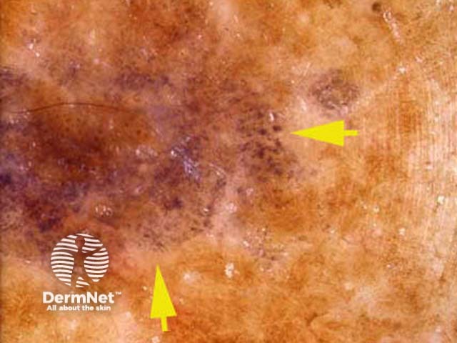
Lichenoid inflammation affecting a solar lentigo or seborrhoeic keratosis typically results in localised destruction of melanocytes and free melanin in the dermis or melanin within melanophages. These appear as granular areas of grey dots. Grey dots can also be typically seen within melanoma. However the lichenoid keratosis has no pigment network and there are usually amorphous areas with or without keratinous surface /or other features of seborrhoeic keratosis.


Porokeratosis is distinguished by a cornoid lamella around the lesion. Sometimes there is prominent follicular plugging


Sebaceous hyperplasia is distinguished by pale yellow lobules around a central follicular opening. Telangiectasia is common but tends to be uniform, in contrast to the irregular arborising vessels seen in basal cell carcinoma.


Viral warts are keratinocytic lesions with a lobular structure (like frog spawn), sometimes with a central thrombosed capillary within each lobule. The normal dermatoglyphics are interrupted. Some have a papilliform structure.
In contrast, a corn has a translucent central core, and a callus is hyperkeratotic without other distinguishing features.


An epidermal naevus resembles a seborrhoeic keratosis or viral wart, with fissures, crypts and milia. However it is very uniform in appearance and appears within the first decade.


The areola and nipple are usually clinically obvious of course. However, an accessory nipple (present in 1 in 18 individuals) may resemble a compound naevus. Characteristically, the breast tissue has a delicate uniform peripheral pigment network.


Close inspection of a cyst will show the follicular opening.


The clear cell acanthoma is an unusual benign epidermal tumour with characteristic dermoscopic features. There are multiple pinpoint or dotted vessels arranged in line like a string of pearls.


Trichoepithelioma and trichadenoma are benign hair follicle tumours. Dermoscopy of these lesions usually shows multiple white clods of variable diameter ( milia like cysts ). This allows them to be distinguished from basal cell carcinoma on most occasions.


*Images supplied by Dr Mike Inskip
The vascular pattern seen on dermoscopy can be used to diagnose red scaly plaques:


Entodermoscopy is the use of dermatoscopy to:


A dermatoscope can be used to evaluate capillaries. For example


How does nail fold capillaroscopy distinguish lupus erythematosus from systemic sclerosis?
See the DermNet bookstore.

