DermNet provides Google Translate, a free machine translation service. Note that this may not provide an exact translation in all languages
Cutaneous mastocytosis images
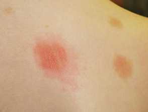
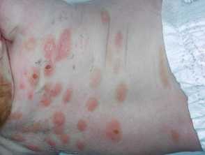
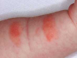
A positive Darier sign - rubbing an area of mastocytosis has resulted in redness, swelling, and urtication in 5 minutes
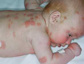
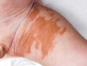
Extensive infantile mastocytosis - the yellow-orange colour and positive Darier sign is diagnostic
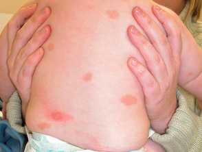
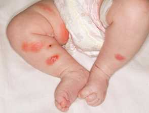
Mastocytosis in a baby - red-brown patches are characteristic. The left lower back lesion has urticated after rubbing - a positive Darier sign
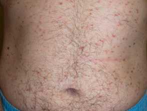
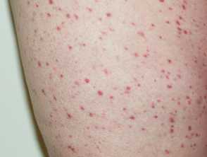
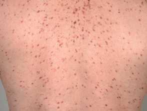
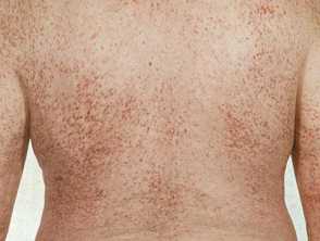
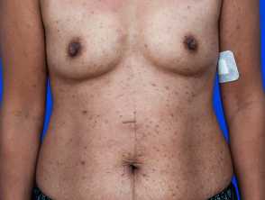
Red-brown maculopapules typical of adult-onset urticaria pigmentosa
(CM-patient1)
Red-brown papules of adult-onset urticaria pigmentosa on the abdomen
(CM-patient1)
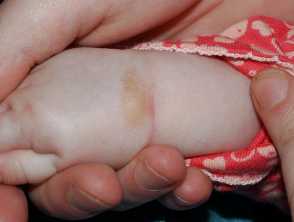
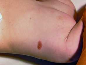
A solitary mastocytoma on the wrist - it will urticate if rubbed
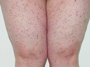
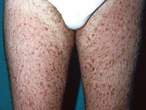
Telangiectasia macularis eruptiva perstans - the lesions are more red in this variant of mastocytosis
Adult-onset urticaria pigmentosa - the lesions are more red than usual, and are often brown in colour
(CM-patient2)
Adult-onset urticaria pigmentosa - the lesions are more red than usual, and are often brown in colour
(CM-patient2)
Yellow-brown pigmented macules in juvenile mastocytosis
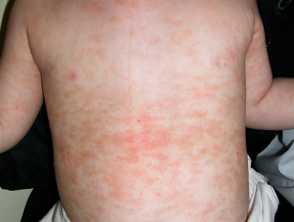
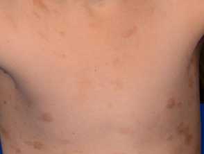
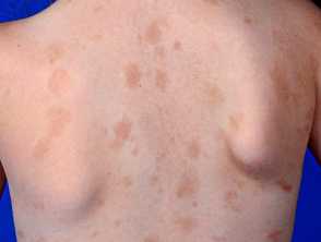
Juvenile mastocytosis: red-brown macular lesions on the trunk. They are likley to resolve over the next few years
(CM-patient3)
Juvenile mastocytosis: red-brown macular lesions on the trunk. They are likely to resolve over the next few years
(CM-patient3)
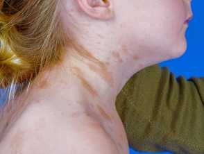
Juvenile mastocytosis: red-brown macular lesions on the neck. They are likely to resolve over the next few years
(CM-patient3)
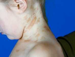
Juvenile mastocytosis: red-brown macular lesions on the neck. They are likely to resolve over the next few years
(CM-patient3)
A solitary mastocytoma on an infant's chest
Red-brown monomorphic maculopapules in adult-onset urticaria pigmentosa
Red-brown monomorphic maculopapules in adult-onset urticaria pigmentosa
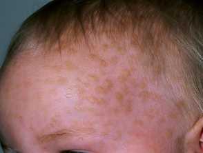
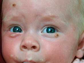
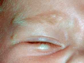
Lesions of infantile mastocytosis on the face and lids
(CM-patient4)
Infantile mastocytosis - some lesions have blistered after urticating due to rubbing
(CM-patient4)
Infantile mastocytosis - crusting is evident where blistering has been present
(CM-patient4)
Infantile mastocytosis - crusting is evident where blistering has been present
(CM-patient4)
Urticated plaques on the arm due to infantile mastocytosis
(CM-patient4)
Lesions of mastocytosis on the lid
(CM-patient4)
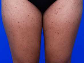
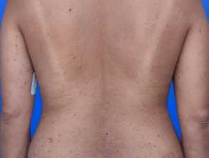
Adult-onset urticaria pigmentosa in skin of colour
(CM-patient5)
Adult-onset urticaria pigmentosa in skin of colour
(CM-patient5)
Adult-onset urticaria pigmentosa in skin of colour
(CM-patient5)
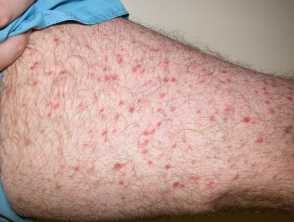
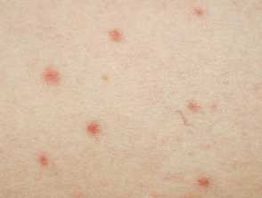
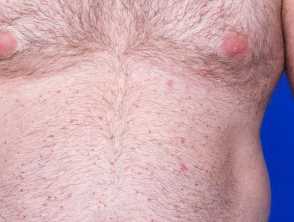
Red monomorphic maculopapules due to a variant of mastocytosis called telangiectasia macularis eruptiva perstans
(CM-patient6)
Urticaria pigmentosa in an adult - dermoscopy showed telangiectasia; so called telangiectasia macularis eruptiva perstans
(CM-patient6)
Red-brown maculopapules in adult onset urticaria pigmentosa
Diffuse cutaneous mastocytosis in an infant - the tan-orange colour is typical; blistering is more common in this variant