Introduction Demographics Causes Clinical features Complications Diagnosis Treatment
Pilomatricoma is an uncommon, harmless, hair follicle tumour derived from hair matrix cells. It is also spelled ‘pilomatrixoma’, and sometimes known as ‘calcifying epithelioma of Malherbe’.
Pilomatricoma is most often diagnosed in young children but may also affect adults. It appears to be slightly more common in females than males.
Cases have been reported of multiple pilomatricomas in association with the rare neurological condition myotonic dystrophy. Individual cases have also been reported of pilomatricomas arising in patients with a variety of other genetic disorders. The vast majority are not associated with any other abnormality.
The cause of pilomatricoma is now known to be due to a localised mutation in a hair matrix cell. An overactive proto-oncogene called BCL-2 suggests the normal process of cell death is suppressed, and mutations in CTNNB1 in most cases suggest loss of regulation of a protein complex called beta-catenin/LEF-1.
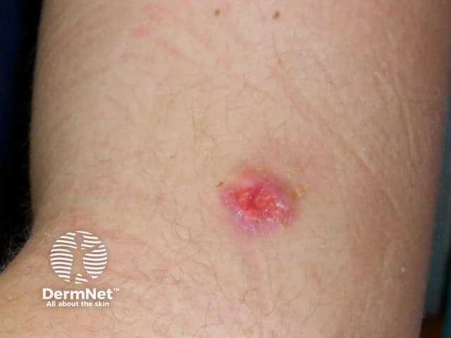
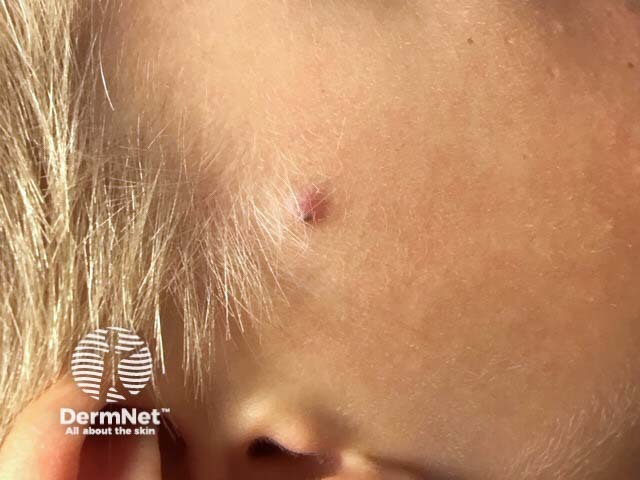
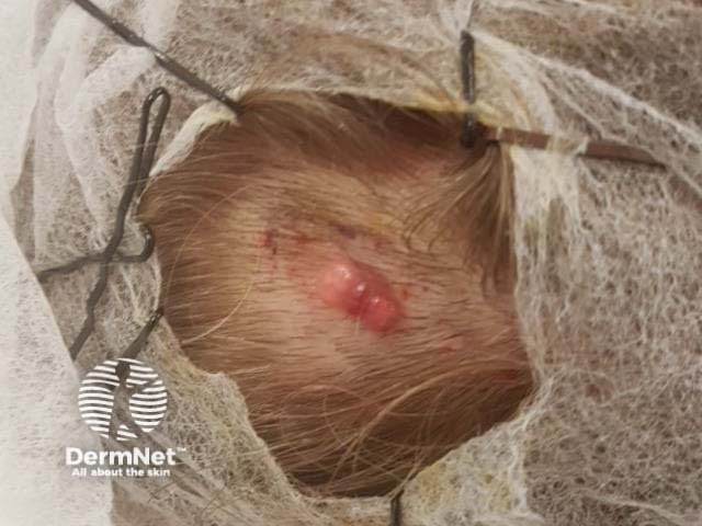
Pilomatricoma presents as a single skin-coloured or purplish lesion.


Complications of pilomatricoma are rare. However, occasionally they grow to giant size (several centimetres in diameter), and pilomatrix carcinoma (cancer) has been very rarely reported.
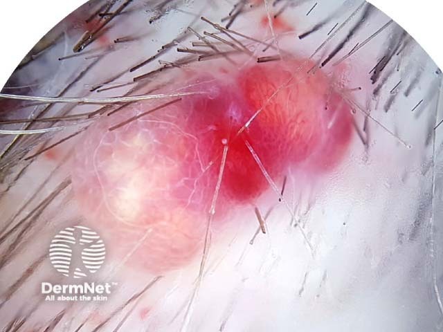
If pilomatricoma is suspected, dermoscopy may be helpful, showing a central whitish or grayish-blue structureless area. Erythema and telangiectasia are sometimes observed.

If the nature of the skin lesions is uncertain, ultrasound scan may be recommended. The scan of pilomatricoma is described as a doughnut within the dermis (mid layer of the skin) with a tail (the tail denotes calcification). Alternatively, the calcification may be detected by X-ray.
A biopsy will help to establish the cause of the lesion. Alternatively the whole lesion can be removed, providing both diagnosis and treatment. The histology of pilomatricoma is striking. It may show a sharply demarcated tumour surrounded by a fibrous capsule or a poorly demarcated tumour without capsule. There are darkly stained ‘basophilic’ cells and ‘shadow’ cells with missing nuclei. Calcium deposits are found in most lesions.
Pilomatricomas are usually surgically excised. They do not disappear by themselves, and if incompletely removed, they may recur.

