What is an angiokeratoma?
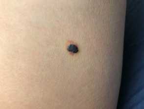
An angiokeratoma is a benign capillary ectasia in the superficial dermis that presents as an asymptomatic blue-red hyperkeratotic papule anywhere on the skin.
Angiokeratomas
Who gets angiokeratomas and how are they classified?
Table 1. Classification of angiokeratomas
| Who gets it? | Clinical/Distribution | |
| Sporadic angiokeratoma | Common, increases with age | Anywhere on the skin, sometimes on mucous membranes |
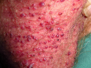
| Angiokeratoma of Fordyce | Common, male predominance, increases with age | Found on scrotum and vulva |
| Angiokeratoma of Mibelli | Rare, appears in childhood | Multiple warty purple papules; acral, elbows, knees, breast |
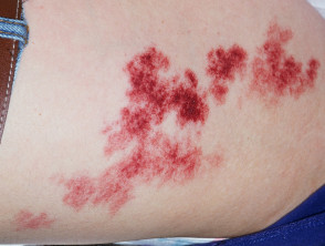
| Angiokeratoma circumscriptum | Rare vascular birthmark, female predominance | Cluster of angiokeratomas |
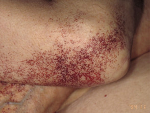
| Angiokeratoma corporis diffusum (Fabry disease) | Rare X-linked genetic disorder, more severe in males, milder in carrier females | Angiokeratomas most numerous over buttocks, groin, and lower trunk |
| Acral pseudolymphomatous angiokeratoma of children (APACHE) | Rare, predominantly seen in children | Red-purple angiokeratoma-like papules on arms and legs, characteristic histology |
| Drug-induced eruptive angiokeratomas | Rare drug side effect – heparin (enoxaparin) | Multiple eruptive lesions |
| Penile angiokeratomas (PEAKERs) | Subtype of genital angiokeratomas in males including children | Glans penis and penile shaft, 20% associated with scrotal lesions |
Angiokeratomas are one of the multiple vascular lesion types described in Cobb syndrome.
What are the clinical features of angiokeratoma?
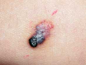
Angiokeratomas are red-purple papules seldom more than 5 mm in diameter with a smooth or roughened surface.
Angiokeratomas
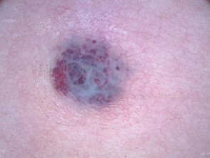
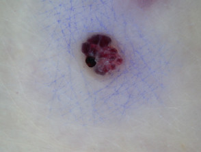
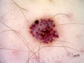
Dermoscopy of angiokeratoma
- Dark lacunae due to the dilated capillaries in the papillary dermis
- Whitish veil due to the hyperkeratosis
Dermoscopy of angiokeratomas
[see more Angiokeratoma images]
How do clinical features vary in differing types of skin?
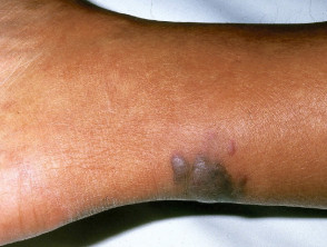
Angiokeratomas are less obvious in skin of dark colour.
What are the complications of angiokeratoma?
- Bleeding
- Thrombosis resulting in inflammation and pain
How is angiokeratoma diagnosed?
Angiokeratoma is usually diagnosed clinically on the typical appearance but can be confirmed on dermoscopy and skin biopsy [see Angiokeratoma pathology].
If Fabry disease is suspected, a blood test in males should be undertaken for alpha-galactosidase A activity. Genetic analysis (GLA gene) of affected males and carrier females will confirm the diagnosis. Skin biopsy may show intracellular accumulation of globotriasylceramide (Gb3).
APACHE is diagnosed on histology with characteristic lymphoid hyperplasia and proliferation of thick-walled vessels in the upper dermis, and is not a true angiokeratoma despite the clinical resemblance.
What is the differential diagnosis for angiokeratoma?
What is the treatment for angiokeratoma?
Angiokeratomas do not usually require treatment. Excisional biopsy may be indicated to exclude melanoma in some cases. Cautery, cryotherapy, and vascular lasers may be used to treat bleeding lesions or multiple lesions for cosmetic reasons.
Males with Fabry disease usually require enzyme replacement therapy.
APACHE may respond to topical sirolimus (rapamycin).
What is the outcome for angiokeratoma?
Angiokeratomas do not resolve spontaneously and tend to become more warty with time.