Introduction
Botryomycosis (bacterial pseudomycosis) is an uncommon chronic bacterial infection of skin caused by masses of colonizing bacteria. Staphylococcus aureus is the usual cause, but a wide range of gram negative organisms have also been isolated.
Histology of botryomycosis
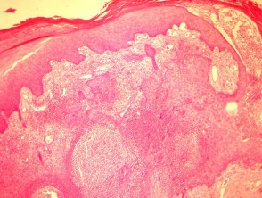
Histopathologic examination of botryomycosis shows a marked inflammatory response, scarring and suppuration. Trans-epidermal elimination, and marked epithelial hyperplasia may also sometimes be seen (figure 1).
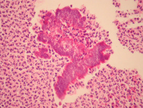
Botryomycosis resembles mycetoma with granules present within suppurative inflammation. The granules are composed of blue-stained bacteria surrounded by an intensely eosinophilic coat (the Splendore-Hoeppli Phenomenon, figure 2).
Botryomycosis pathology
Special stains for botryomycosis
Gram stain will detect bacterial forms of botryomcyosis. PAS stain stains the granule.
Differential diagnosis of botryomycosis pathology
Mycetoma – In contrast to mycetoma, botryomycosis is not caused by fungi or actinomycosis.
Disseminated infections – A disseminated infection will have a diffuse pattern of invasion without localized granules.
Squamous cell carcinoma – A massive epidermal reaction (figure 1) can be confused with an invasive carcinoma, particularly if only the surface of a lesion is biopsied.