What is carcinoma in situ of the oral cavity?
Carcinoma in situ of the oral cavity is also called oral intraepithelial carcinoma and oral squamous cell carcinoma in situ. In carcinoma in situ, cancer cells are confined to the epithelium, in contrast to invasive oral cancer (squamous cell carcinoma, SCC).
Who gets carcinoma in situ of the oral cavity?
Oral intraepithelial carcinoma may affect about 0.5% of the world population, although it is likely to vary with sex, geography and ethnicity.
There is a strong association with tobacco smoking (six times more common in smokers than non-smokers) and alcohol intake (independent of drinking pattern or beverage type). It is also associated with betel quid chewing and oral submucous fibrosis.
It usually appears in adult life with prevalence increasing with increasing age.
- Carcinoma in situ of the oral cavity is found in less than 1% of men under 30 years of age.
- Up to 8% of men over 70 years of age and 2% of women over 70 years of age have carcinoma in situ of the oral cavity.
- It is rare before age 30 and peaks after 50 years.
- It mainly affects middle-aged to elderly men
- Non-smokers are likely to present at an older age.
What are the clinical features of carcinoma in situ of the oral cavity?
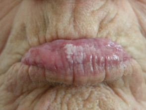
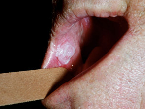
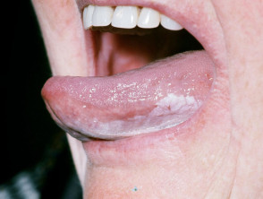
Early carcinoma in situ of the oral cavity is a slightly elevated grey-white plaque either well defined or which blends in gradually with surrounding mucosa. It can be a localised solitary lesion or multifocal and diffuse.
Two clinical forms are recognised.
1. Homogeneous — refers to homogeneous uniform colour AND texture
- Uniform white colour (before diagnosis, this may be termed leukoplakia)
- Uniform flat, thin appearance
The surface may become leathery — smooth, wrinkled, corrugated or with shallow cracks. This form is usually asymptomatic.
2. Non-homogeneous — refers to an irregularity of either the colour OR the texture
- Predominantly white or white-red (before diagnosis, this may be termed erythroleukoplakia)
- An irregular surface which can be flat, nodular, exophytic, warty
Variants of the non-homogeneous form have been described including nodular, verrucous (including proliferative verrucous) and speckled. This form may be associated with mild discomfort or localised pain.
The most common site for carcinoma in situ of the oral cavity is the inside of the cheeks (the buccal mucosa) and then in decreasing order of frequency:
- Gums (alveolar mucosa)
- Lower lip
- The floor of mouth (under the tongue)
- Sides or undersurface of the tongue (lateral or ventral tongue)
- Soft palate.
Oral leukoplakia
Association with squamous cell carcinoma (SCC)
A large proportion of oral cancers are associated with preceding longstanding carcinoma in situ, especially the proliferative verrucous variant.
There may be no change in appearance or symptoms in the early stages of cancer development. Classic changes of cancer are ulceration, induration/hardness, bleeding and tumour outgrowth.
Risk factors for the development of SCC development
- Dysplasia (atypical changes) on histology is regarded as the most important factor. However, dysplastic lesions can resolve spontaneously, and nondysplastic lesions may develop into cancer.
- Site — the floor of the mouth under the tongue and the sides/undersurface of the tongue
- Clinical type — speckled non-homogeneous, especially proliferative verrucous leukoplakia
- Female sex
- If the preceding carcinoma in situ is NOT associated with tobacco use.
- A long duration of carcinoma in situ
- Large lesion size
- Presence of Candida albicans — note that this is most commonly found in lesions at the angles of the mouth or top surface of the tongue, which are rare sites for cancer development.
No molecular tumour markers have yet been found that can be used to predict cancer development in an individual or lesion. The role of human papillomaviruses (wart virus) has not yet been determined.
How is the diagnosis made?
- Biopsy of clinically suspected oral leukoplakia is mandatory to: exclude recognised diseases, and to assess for the absence or presence and grade of dysplasia.
- It is appropriate to wait two weeks after the first presentation to assess clinical response to initial treatment, for example, for candida, change in tooth brushing habit, cessation of smoking, etc.
- The biopsy may be incisional or excisional, single or multiple and may be done under local or general anaesthetic depending on site, the number of biopsies required and the type of biopsy.
- Biopsies should be taken from either a symptomatic area or if asymptomatic then from red or indurated areas.
- The presence of dysplasia, carcinoma-in-situ and invasive carcinoma cannot always be predicted clinically.
The histopathology of oral leukoplakia is not always diagnostic. Epithelial changes range from atrophy (thinned) to hyperplasia (thickened) and it may show hyperkeratosis. Dysplasia (atypical changes) may be mild, moderate, severe, carcinoma in situ or invasive carcinoma. The pathology report must comment on the absence or presence of dysplasia, and the severity.
Treatment of oral carcinoma in situ
It is not known if early active treatment of oral squamous cell carcinoma in situ prevents the development of invasive squamous cell carcinoma. There is a high recurrence rate after treatment.
- Avoid aggravating habits, for example, quit smoking.
- Surgical excision
- CO2 laser — excision or vaporisation
- Oher options include retinoids (acitretin or isotretinoin), photodynamic therapy.
Lifelong follow-up is recommended whether or not the disorder has been treated:
- 3-12 monthly clinical checks
- Biopsy of suspicious changes
The oral mucosal examination must include the floor of mouth and sides of tongue using gauze to hold the tip of the tongue and pull upwards and side to side. Most oral SCC develops in the sides and undersurface of the tongue, the floor of the mouth and back to the soft palate and tonsillar area.


