What is lichen sclerosus?
Lichen sclerosus is a chronic inflammatory skin condition with sclerotic and atrophic lesions of the anogenital area and extragenital skin.
What are the clinical features of lichen sclerosus?
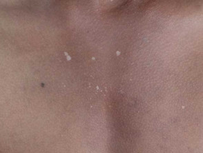
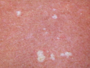
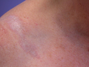
Lichen sclerosus presents as distinctive porcelain white crinkled or thickened patches that tend to scar.
Clinical features of cutaneous lichen sclerosus
See also Extragenital lichen sclerosus images, Images of penile lichen sclerosus, Perianal lichen sclerosus images, and Vulval lichen sclerosus images.
What are the dermoscopic features of lichen sclerosus?
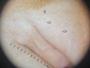
Extragenital cutaneous lichen sclerosus
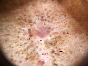
Dermoscopy of cutaneous lichen sclerosus varies with the age of the lesion.
Early lesion (less than 2 years):
- White, yellowish, or pale brown follicular plugs (‘comedo-like openings’)
- Homogeneous structureless white to pinkish white background
- Vessels may be linear or dotted
- Blue-grey peppered dots and globules.
Late lesion (more than 2 years):
- Comedo-like openings are less frequent in older lesions
- Poorly defined bright white or red structureless areas with a diffuse margin lacking the red halo
- White shiny streaks
- Linear curved or branching vessels are less common than in anogenital lichen sclerosus
- Dotted, commalike, and hairpin vessels may be seen
- If itchy, there may be purple dots
- Scattered blue-grey dots
- Pigment network in sclerotic lesions.
Dermoscopy of cutaneous extragenital lichen sclerosus
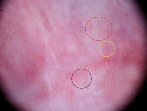
Genital lichen sclerosus
Dermoscopy of genital lichen sclerosus shows:
- A whitish background
- Patchy structureless areas varying from white, to white-yellow, or milky-pink
- Linear and (more commonly) dotted vessels, but markedly decreased compared to unaffected surrounding skin
- Peppered blue-grey dots and globules can be so marked as to mimic melanoma
- Purpuric red-purple globules and blotches due to scratching.
Dermoscopy features of genital lichen sclerosus
What is the dermoscopic differential diagnosis for lichen sclerosus?
Vitiligo dermoscopy: white structureless areas described as having a ‘white glow’ due to the lack of pigment network.
Idiopathic guttate hypomelanosis dermoscopy: white structureless ‘cloudy sky’ areas in various patterns such as amoeboid, petaloid, feathery, and nebuloid.
Morphoea dermoscopy: fibrotic beams (’white clouds’), pigment network, and spreading telangiectasia.
What is the histological explanation of the dermoscopic features of lichen sclerosus?
- White areas and scale — epidermal atrophy and hyperkeratosis
- Comedo-like openings — follicular plugging
- Structureless areas — dermal sclerosis and homogenisation
- White shiny streaks — increased dermal collagen
- Blue-grey peppering — dermal melanophages of post-inflammatory hyperpigmentation
- Pigment network — epidermal hyperpigmentation
- Purpuric red-purple globules are blood spots/haemorrhage from scratching.