DermNet provides Google Translate, a free machine translation service. Note that this may not provide an exact translation in all languages

Translate

Ocular melanoma

Author: Dr Giri Raj, Dermatologist, New Plymouth, New Zealand; Dr Amanda Oakley, Dermatologist, Hamilton, New Zealand, 2013.


toc-icon

What is ocular melanoma?

Melanoma is a malignant tumour of melanocytes. Ocular melanoma refers to melanoma of the eye, and accounts for about 5% of all melanomas [1]. Primary ocular melanoma is the most common primary malignant tumour of the eye in adults [2].

Normal anatomy of the eye

  • Conjunctiva – the membrane on the outer aspect of the eye.
  • Sclera – the white of the eye, an opaque fibrous protective layer around the eye. It is continuous with the cornea.
  • Cornea – the clear front surface of the eye.
  • Iris – the coloured part of the eye. It can control the amount of light entering the eye by opening and closing.
  • Ciliary body – a muscle that enables the eye to focus by changing the shape of the lens.
  • Choroid – a layer of tissue in the back of the eye between the sclera and the retina.

Melanocytes

Melanocytes are cells that produce pigment (melanin). Dark skin has more active melanocytes than has fair skin. More melanin is produced by melanocytes in response to sun exposure and may result in patchy pigmentation on the surface of the eye, called melanosis.

Melanocytic naevi

Melanocytes often cluster as pigmented moles on the skin, properly called melanocytic naevi. They are harmless. Melanocytic naevi may also arise on the conjunctiva. They are sometimes present at birth (congenital melanocytic naevus).

Collections of melanocytes in the deeper tissues of the eye are called ocular melanocytosis. Naevus of Ota is a form of dermal melanocytosis that may involve the eye.

What parts of the eye are affected by melanoma?

Primary ocular melanomas can be divided into 2 types:

  • Uveal melanomas: these arise within the iris, choroid and ciliary body. They are the most common form of ocular melanoma, with a reported stable incidence of 7 per million per year in the USA. The majority of cases (over 90%) involve the choroid. Approximately 6% involve the ciliary body and 4% involve the iris.
  • Conjunctival melanomas. They are less frequent though the incidence is increasing. Conjunctival melanomas have an incidence of less than 5 cases per million per year.

Uveal melanoma seen through slit lamp* (4 of choiroid, 1 of iris)

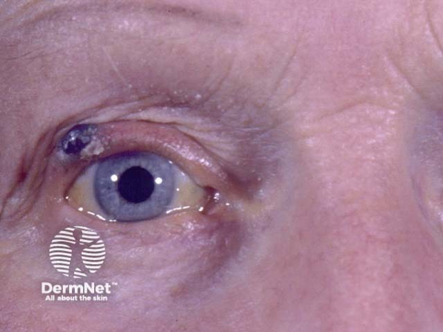
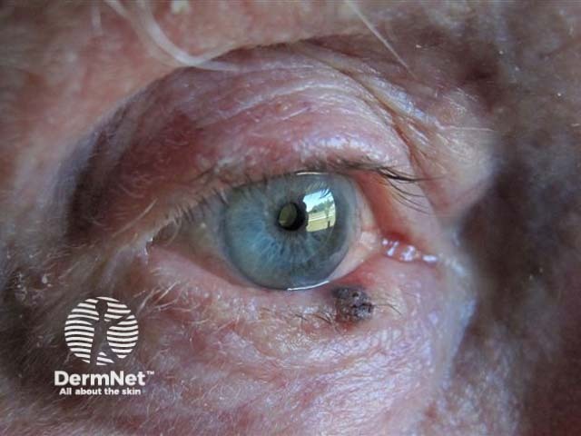
*Images supplied by Dr Stephen Guest

Cutaneous melanoma can also affect the eyelid, but these are not considered ocular melanomas.

Melanoma affecting eyelid**

**Images supplied by Dr Stephen Ng

What is the cause of ocular melanoma?

Like other forms of melanoma, ocular melanoma occurs because of genetic changes within melanocytes that cause the cells to proliferate. Further changes in the cells cause them to invade surrounding tissues and to spread elsewhere round the body (metastasise).

Many reports suggest exposure to sunlight may be an important factor in the development of ocular melanoma.

Who gets ocular melanoma?

Both males and females appear to have equal incidence of ocular melanoma. The peak incidence of presentation is closest to 62 years of age.

Those most at risk of ocular melanoma are of Caucasian race and have fair skin and light iris colour. However, melanoma may also affect those with darker skin and eye colour.

Melanosis, congenital ocular melanocytosis and neurofibromatosis are also associated with increased risk.

Patients with conjunctival melanoma tend to have many melanocytic naevi (moles). It is thought that about 20% arise from naevi and about 60-75% within conjunctival melanosis.

How is the diagnosis of ocular melanoma made?

Diagnosis of ocular melanoma is usually made by accurate clinical examination. The main indicators of disease in 90 patients described in a Canadian study were:[3]

  • partial loss of visual field (in 33% of patients)
  • sun sensitivity (in 20%)
  • blurred vision (in 20%)
  • incidental finding on eye examination (in 17%)

Conjunctival melanoma presents as an increasingly irregular pigmented lesion on the external eye.

Other symptoms may include a protruding eye, change in colour of the iris, red or painful eye, and retinal detachment.

Progression of ocular melanoma

Half of all patients develop metastatic disease within 15 years after the primary tumour has been treated. Unfortunately there is no cure for metastatic disease.

Metastasis occurs by either local extension or blood dissemination. Transscleral spread may occur through ciliary nerves, veins, arteries or aqueous drainage channels. There is no lymphatic network in the uveal tract. Distal metastases occur in the liver in over 80% of cases. The lung, bones and skin are sometimes also involved.

Poor prognosis is associated with:

  • large tumour size
  • ciliary body involvement
  • older patient age.

When treated early, melanoma of the iris is less likely to impair vision or metastasise when compared to other uveal melanomas.

Histological features associated with higher metastatic risk include:

  • the presence of cells with epitheloid morphology
  • increased infiltration with immune cells
  • high mitotic rate
  • increased microvascular density
  • loss of BRCA–associated protein 1 staining.

Chromosomal changes in ocular melanoma

Advances in detecting genetic changes in melanoma enables the diagnosis of tumours with high metastatic potential.

  • BRAF mutations are frequent in iris melanomas (like cutaneous melanomas) but are extremely rare in other types of uveal melanoma.
  • GNA11 or GNAQ mutations are found in 80-85% of uveal melanomas and are early events in development of non-cutaneous melanoma. The presence of these mutations are not prognostic for later metastatic potential. GNAQ/GNA11 mutations are also seen in ocular melanocytosis, blue naevus and naevus of Ota.[4,5]
  • Mutations in BRCA1 associated protein-1 (BAP-1) and splicing factor 3B subunit 1 (SF3B1) occur later in disease progression. The presence of BAP-1 mutations are strongly associated with metastatic disease. A more favourable outcome is associated with presence of SF3B1 mutations.

Genetic alterations associated with poorer prognosis in uveal melanoma include:

Chromosome 6p gain is associated with a better prognosis and it may act to delay or prevent chromosome 3 loss.

What tests are done in ocular melanoma?

Which tests are chosen depends on the clinical presentation and what is available. They may include:

  • Fundus imaging / photography
  • Ultrasound scan of the eye
  • Biopsy of the tumour
  • CT scan of the eye and brain
  • Liver ultrasound scan
  • PET-CT scan.

What is the treatment for ocular melanoma?

Local treatments for ocular melanoma have improved, with increasing preservation of normal eye tissue, however survival rates remain unchanged.

Uveal melanoma

Plaque radiotherapy

  • Radioactive seeds (most often Iodine-125) contained within a small disc or plaque are placed onto the outer aspect of the eye overlying the tumour. After a few days the plaque is removed.

Surgical resection

  • Iridectomy
  • Choroidectomy
  • Enucleation (removal of the affected eye) if tumours i thicker than 8 mm or orbital spread is extensive

Other

  • Phototherapy/ transpupillary thermotherapy: infrared lasers heat the tumour by a few degrees to disrupt cellular metabolism. This treatment is often used alongside radiotherapy to reduce the incidence of local recurrence.
  • External proton beam radiotherapy may give high rates of local control, however significant loss of vision may also occur.
  • Gamma knife stereotactic radiosurgery.

Conjunctival melanoma

Conjunctival melanoma is treated by:

  • wide local excision, and;
  • adjuvant therapy, such as brachytherapy, cryotherapy, or application of a topical chemotherapeutic agent.

Metastatic ocular melanoma

Prognosis in patients with metastatic disease remains poor. Options for metastases from ocular melanoma may include:

  • Chemotherapy using melphalan injected directly into liver metastases
  • Surgical removal of the affected part of liver
  • Ipilumumab immunotherapy. This monoclonal antibody blocks an antigen on cytotoxic T lymphocytes so that the immune system can target melanoma cells.
  • Selective internal radiation therapy, which involves infusion of radioactive yttrium beads into the liver, where they accumulate within melanoma cells.

What happens at follow-up of ocular melanoma?

After treatment of the primary tumour, regular follow-up is important to check for local recurrence or systemic spread.

As the liver is the most common first site of systemic spread, it is often the target of follow-up investigations such as liver function tests, abdominal ultrasound scan and abdominal magnetic resonance imaging (MRI).

What is the outlook for patients with ocular melanoma?

Unfortunately metastatic melanoma remains the leading cause of death among patients with ocular melanoma. The extent of systemic spread and tumour burden determines the average length of survival after liver metastases have been detected.


References

  1. Dermatology. Eds. Bolognia JJ, Jorizzo JL, Schaffer JV. 3rd edition 2012.
  2. Jovanovic P, Mihajlovic M, Djordjevic-Jocic J, Vlajkovic S, Cekic S, Stefanovic V. Ocular melanoma: an overview of the current status. Int J Clin Exp Pathol. 2013 Jun 15;6(7):1230-44. Print 2013. PubMed PMID: 23826405; PubMed Central PMCID: PMC3693189. PubMed
  3. Gallagher RP, Elwood JM, Rootman J, Threlfall WJ, Davis J. Symptoms and time to presentation and treatment in ocular melanoma: the Western Canada Melanoma Study. Can J Ophthalmol. 1988 Feb;23(1):11–3. PubMed PMID: 3349359.
  4. Eagle RC Jr. The pathology of ocular cancer. Eye (Lond). 2013 Feb;27(2):128-36. doi: 10.1038/eye.2012.237. Epub 2012 Nov 16. Review. PubMed PMID: 23154492; PubMed Central PMCID: PMC3574240.
  5. Mudhar HS, Doherty R, Salawu A, Sisley K, Rennie IG. Immunohistochemical and molecular pathology of ocular uveal melanocytoma: evidence for somatic GNAQ mutations. Br J Ophthalmol. 2013 Jul;97(7):924–8. doi: 10.1136/bjophthalmol-2013-303291. Epub 2013 May 17. PubMed PMID: 23685997. PubMed

On DermNet

Other websites

Books about skin diseases

Text: Miiskin


Related information

Sign up to the newsletter