What is onychomatricoma?
Onychomatricoma is a rare, benign fibroepithelial tumour of the nail matrix [1].
Onychomatricoma
Who gets onychomatricoma?
Onychomatricoma typically affects Caucasian women, with a peak incidence in the fifth decade of life. It is rarely observed in children [2].
What causes onychomatricoma?
The exact pathophysiology of onychomatricoma is unknown, although it could be precipitated by trauma [3,4].
What are the clinical features of onychomatricoma?
Onychomatricoma more commonly affects the fingers than the toes. It could involve either a single digit or multiple digits simultaneously [2,5].
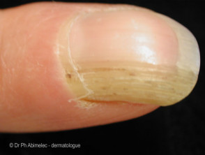
Onychomatricoma is typically slow-growing and painless. Its classic clinical features include [1,4,5]:
- A yellowish and thickened nail plate
- Proximal splinter haemorrhage
- Longitudinal overcurvature of the nail plate
- Finger-like projections penetrating the nail plate, resulting in the characteristic 'woodworm' cavities when viewed from the nail plate’s free margin.
Rarer presentations of onychomatricoma include longitudinal melanonychia, subungual haematoma, and dorsal pterygium [4,6].
Dermoscopic features of onychomatricoma include perforations in the distal portion of the nail plate, haemorrhagic striae, as well as white longitudinal grooves corresponding to the nail plate channels [5].
What are the complications of onychomatricoma?
Onychomatricoma may be complicated by onychomycosis, so both conditions can coexist [1,5]. Delayed diagnosis and treatment are not uncommon due to unfamiliarity with onychomatricoma [2].
How is onychomatricoma diagnosed?
Onychomatricoma is diagnosed based on its characteristic clinical features supplemented by dermoscopy, imaging, and histopathology [6].
Imaging may be considered to guide clinical decisions before excision if the presentation is non-specific or unclear [4]. Imaging modalities that could help with diagnosing onychomatricoma include [5,7]:
- Ultrasound — this may show hypoechogenic tumour affecting the nail matrix and hyperechogenic area corresponding to the finger-like projections; reduced blood flow could also be observed
- MRI — the affected nail matrix typically shows a low signal uptake, while the finger-like projections have high uptake.
Histopathological evaluation could be performed on either nail clippings or a resected tumour, which is considered the gold standard [1,4–7]:
- Nail clippings — these classically show a thickened nail plate with cavities filled with serous material, lined by a thin layer of epithelium at the periphery
- A resected tumour — distinctive histological features confirm the diagnosis.
- The proximal nail folds — ungual protrusions and deep epithelial invaginations
- The distal zone (corresponding to the lunula) — fibroepithelial projections perforating the nail plate, originating from the matrix.
Immunohistochemistry although helpful, is not routinely required in typical cases [5].
What is the differential diagnosis for onychomatricoma?
Common differential diagnoses of onychomatricoma include:
- Fibrokeratoma (a rare neoplasm)
- Periungual fibroma
- Onychomycosis
- Squamous cell carcinoma
- Intraepidermal squamous cell carcinoma
- Subungual viral wart.
What is the treatment for onychomatricoma?
The definitive treatment for onychomatricoma is complete surgical excision [5].
What is the outcome for onychomatricoma?
Onychomatricoma typically resolves without local recurrence with complete surgical excision [2,4].