What are perpendicular white lines?
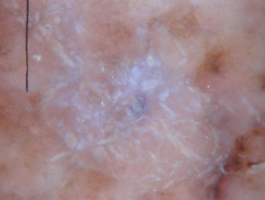
In dermoscopy, perpendicular white lines are short discrete white lines oriented parallel and orthogonal (perpendicular) to each other and seen only under polarised light [1]. They are also known as polarising white lines, short white lines, shiny white lines, shiny white streaks, chrysalis, chrysalids, and crystalline structures. Perpendicular white lines are a clue to a specific diagnosis including basal cell carcinoma (BCC) and some melanomas [2].
What do perpendicular white lines look like through the dermatoscope?
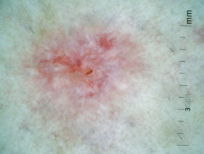
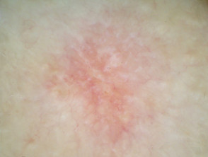
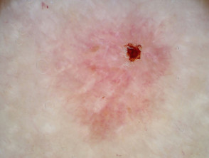
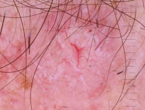
Perpendicular white lines are only seen under polarised light. They appear as short, shiny white lines and move as the dermoscopy lens is moved at different angles over the lesion.
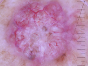
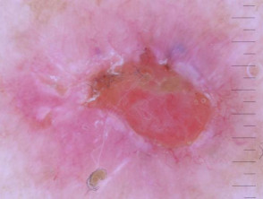
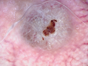
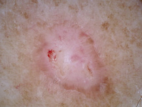
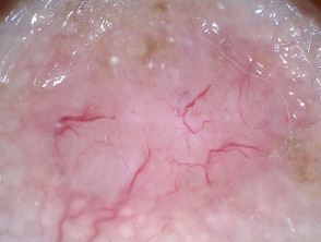
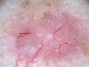
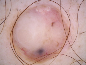
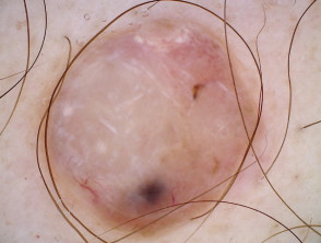
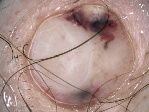
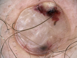
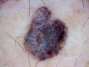
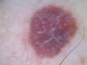
Perpendicular white lines in BCCs
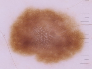
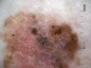
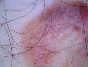
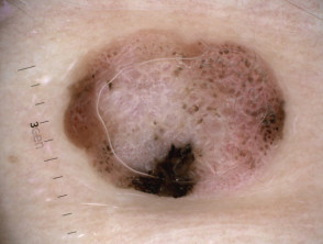
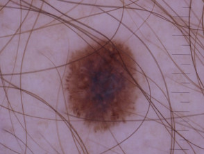
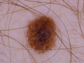
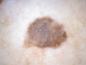
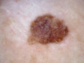
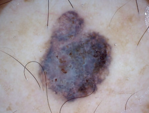
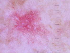
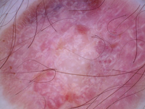
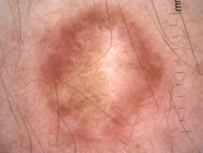
Perpendicular white lines in melanoma
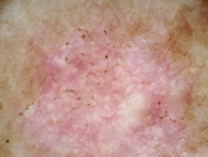
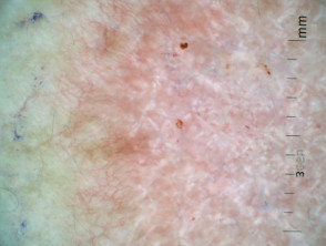
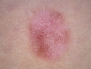
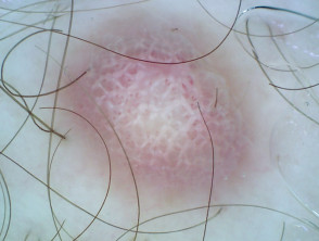
Polarised and non-polarised light
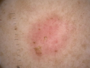
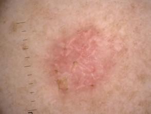
The following pairs of images demonstrate the differences seen in dermoscopy of perpendicular white lines under polarised and nonpolarised light.
Polarised and nonpolarised light in BCC
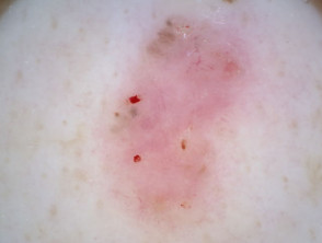
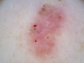

Polarised and nonpolarised light in melanoma
In which lesions are perpendicular white lines seen in through the dermatoscope?
Perpendicular white lines can be seen in the following lesions:
- Pigmented and nonpigmented basal cell carcinoma
- Melanoma
- Spitz naevus
- Dermatofibroma (the polarised white lines are often in radial array rather than perpendicular)
- Scar tissue
- Benign lichenoid keratosis.
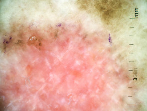
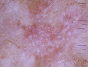
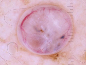
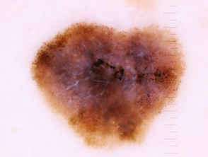
Superficial basal cell carcinoma dermoscopy Nodular basal cell carcinoma dermoscopy Polarised dermoscopy of a nodular basal cell carcinoma presenting as an exophytic polyp Invasive melanoma dermoscopy, Breslow 0.4mm within a melanoma in situ, with associated naevus present Amelanotic melanoma dermoscopy Amelanotic melanoma dermoscopy Spitz naevus dermoscopy Dermatofibroma Dermatofibroma dermoscopyPerpendicular white lines in a variety of lesions
What is the histological explanation of perpendicular white lines?
Perpendicular white lines are thought to correlate histopathologically with altered collagen in the dermis (fibrosis). The birefringent properties of collagen bundles cause rapid randomisation of polarised light. This is the reason collagen appears bright white and is more conspicuous under polarised dermoscopy [3].
They also correlate with dermal invasion in cases of melanoma [4]. However, as our illustrations show, they may also be seen in melanoma in situ.