What is porokeratosis?
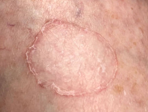
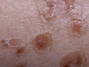
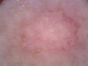
Porokeratosis is a group of skin conditions in which there is abnormal keratinisation. The skin lesions that result are dry and atrophic, with a well-defined ridge-like hyperkeratotic border called a cornoid lamella (best seen on dermoscopy) [1].
Porokeratosis
What are the types of porokeratosis?
Types of porokeratosis include:
- Disseminated superficial actinic porokeratosis
- Porokeratosis of Mibelli
- Linear porokeratosis
- Porokeratosis ptychotropica
- Porokeratosis of Mantoux (also known as porokeratosis palmaris et plantaris disseminata, which is a form of punctate palmoplantar keratoderma in which there are scaly red-brown annular patches on the palms and soles that later spread to the limbs and trunk)
- Porokeratotic eccrine ostial and dermal duct naevus
- Porokeratotic adnexal ostial nevus (PAON)
- Pruritic papular porokeratosis
- Pigmented porokeratosis
- Punctate follicular porokeratosis
Who gets porokeratosis?
Porokeratosis can be present at birth or not develop until adult life [2], depending on the type of porokeratosis.
What causes porokeratosis?
The cornoid lamella in porokeratosis is due to an expanding clonal proliferation of unusual keratinocytes, which is thought to be due genetic mutation [4].
In some types of porokeratosis, a family history is present, consistent with a genetic predisposition [3].
What are the clinical features of porokeratosis?
Each porokeratosis lesion has a characteristic ridge on its border and a central furrow. Their size, onset and distribution depends on the specific type of porokeratosis.
What are the complications of porokeratosis?
The main complication of porokeratosis is a skin cancer, which can develop within a porokeratosis lesion. This may be either a basal cell carcinoma or squamous cell carcinoma, and is more likely to occur in older adults [6]. Porokeratosis should be monitored for malignancy.
How is porokeratosis diagnosed?
The diagnosis of porokeratosis is usually made clinically, often with the help of dermoscopy, but sometimes a biopsy is needed. The biopsy should include the raised edge of the lesion. The pathology of porokeratosis is very distinct, but it may be necessary to point out the clinical features for the pathologist to find a cornoid lamella within the pathological specimen.
Treatment of porokeratosis
There is no known cure for porokeratosis and treatment is generally disappointing. However, the appearance may improve with the following measures:
- 5-fluorouracil cream
- Calcipotriol cream
- Oral acitretin or isotretinoin
- Cryotherapy
- Dermabrasion
- Carbon dioxide laser ablation.
Sun protection is important as exposure to ultraviolet radiation may result in the development of a skin cancer within the linear porokeratosis.