Introduction
Demographics
Causes
Clinical features
Complications
Diagnosis
Differential diagnoses
Treatment
Tinea manuum is a dermatophyte infection of one or both hands. It is much less common than tinea pedis (tinea affecting the foot).
Tinea manuum results from:
It is more likely in those doing manual work, who sweat profusely (hyperhidrosis,) or who already have hand dermatitis.
Zoophilic and geophilic dermatophytes:
Anthropophilic dermatophytes:
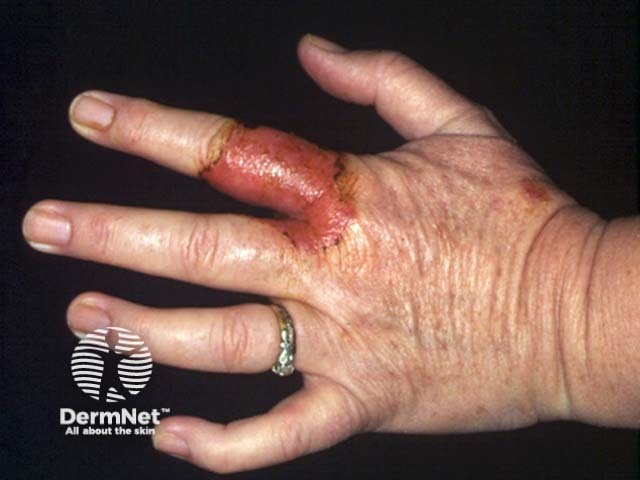
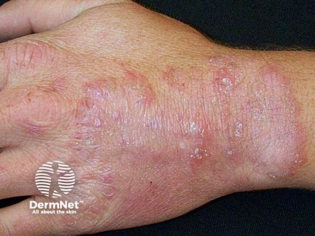
Tinea manuum can occur as an acute inflammatory rash like tinea corporis. There is usually a raised border and clearing in the middle (ringworm). This is most likely when a zoophilic or geophilic fungus is responsible.
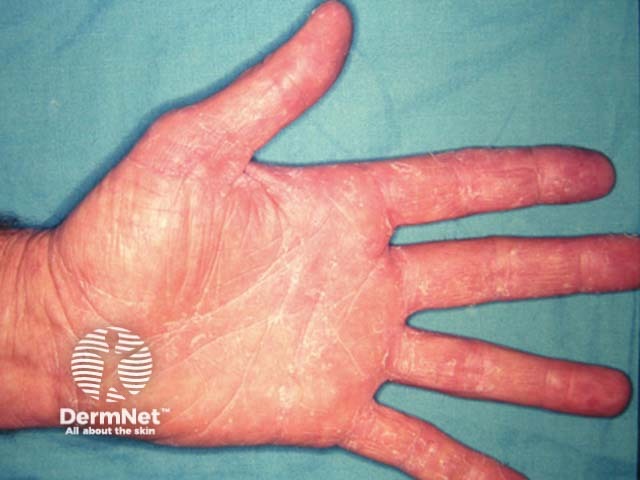
More frequently, tinea manuum causes a slowly extending area of peeling, dryness and mild itching on the palm of one hand (hyperkeratotic tinea). Skin markings may be increased. Generally, both feet appear similar ("one hand, two-foot syndrome"). The usual cause is an anthropophilic (human) fungus:
These fungi may also cause a blistering rash on the edges of the fingers or palm. The blisters appear in crops and contain a sticky clear fluid. They may have a peeling edge. This form of tinea manuum itches and burns.
Tinea manuum can be clinically distinguished from hand dermatitis.
Tinea manuum is a clinical diagnosis confirmed by microscopy and culture of skin scrapings (see Laboratory tests for fungal infection).
Mild tinea manuum is treated with topical antifungal agents, but if the treatment is unsuccessful, oral antifungal medicines may be considered, including terbinafine and itraconazole.

