DermNet provides Google Translate, a free machine translation service. Note that this may not provide an exact translation in all languages

Translate

Quiz
Leg ulceration – 10 cases

This quiz tests your diagnostic skills for leg ulceration.

For each of the ten cases, study the image(s) and then answer the questions. You can click on the image to view a larger version if required.

Each case should take approximately 2 minutes to complete. There is a list of suggested further reading material at the end of the quiz.

When you finish the quiz, you can download a certificate.
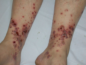
Case 4

A 45-year old man presents with itchy and painful purple plaques, blisters and erosions on his lower legs. He is feeling mildly unwell.

Severe palpable purpura due to acute hypersensitivity vasculitis. Clinically, there are purpuric papules, plaques, haemorrhagic bullae and ulcers.

The differential diagnosis includes IgA-related Henoch-Schönlein purpura and other reasons for purpura including meningococcaemia and hypergammaglobulinaemia. Histologically, there is a small vessel leukocytoclastic vasculitis.

Acute hypersensitivity vasculitis was confined to the skin in this patient, but the gastrointestinal tract, kidneys and joints may also be affected.

Immune complexes, autoantibodies and other inflammatory mediators provoke leukocytoclastic vasculitis. These may arise in response to bacterial or viral infection, drugs (especially antibiotics, NSAIDs and diuretics), collagen vascular disease, inflammatory bowel disease, paraproteinaemia or malignancy (rare). No cause is found in about 50% cases of cutaneous leukocytoclastic vasculitis.

This patient reported that he had a sore throat preceding the eruption. It was believed the vasculitis was due to beta-haemolytic streptococcus, but he had also been treated with a beta lactam antibiotic, which could also be implicated.

In all patients, monitor the extent of internal organ involvement with blood count, ESR, C-reactive protein, renal function, urine microscopy, faecal occult blood, chest X-ray and as otherwise clinically indicated. Most patients should have a skin biopsy to confirm the clinical diagnosis.

Look for a cause: check for hepatitis C, autoantibodies including Rheumatoid Factor and Antinuclear Antibody and cryoglobulins.

Non-specific management should include rest with elevation of the legs and compression. It may require hospital admission – this patient was very reluctant to take time off work!

If a drug is responsible, the vasculitis should clear within about two weeks of discontinuing it. Systemic corticosteroids and/or immunosuppressive agents are not indicated for purely cutaneous leukocytoclastic vasculitis. Colchicine or dapsone may be useful.

The skin eruption settles slowly over some weeks but may recur over a period of months, depending on the underlying cause. Some residual haemosiderin staining is common. This patient had minor scarring at sites of previous ulceration.

Mark question

Sign up to the newsletter