What is Behçet disease?
Behçet disease (BD) is a rare, multi-system condition characterised by recurrent painful oral and genital ulcers, a variety of skin lesions, and eye problems.
Many other organs such as the gut, nervous system, lungs, and arteries may also be affected. It is a form of vasculitis which primarily targets small arteries, but can affect both arteries and veins of all sizes.
It is also known as Adamantiades-Behçet disease, Behçet syndrome, and malignant aphthosis.
Behçet disease
See more Behçet disease images
Who gets Behçet disease?
Behçet disease is most common in individuals originating from eastern and central Asia as well as the eastern Mediterranean. It is most prevalent in Turkey (80–370 cases per 100,000), followed by Iran, and Japan. It is much rarer in those of northern European, African, and Latin American descent.
Onset is usually between 30 and 40 years of age. It rarely occurs in children. It was previously thought that BD was more common in males compared with females, but more recent data suggests that incidence is similar, although this varies between different ethnic and geographic groups.
What causes Behçet disease?
Behçet disease is of unknown cause, but it is thought to have an autoimmune basis. It is associated with several genetic factors, particularly HLA-B51. Several infections have also been implicated as potential triggers including Streptococcus sanguis, herpes simplex virus, hepatitis viruses, and parvovirus B19.
What are the clinical features of Behçet disease?
Common mucocutaneous features
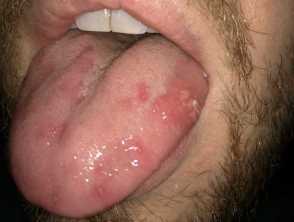
Oral ulcers — anywhere in the mouth, pharynx, and tonsils.
- Typically multiple in number and measure 1–3 cm in size, although they can be larger.
- Usually sharp defined margin with a fibrin-coated base, surrounding erythema, and yellow pseudomembrane.
- Multiple shallow pinpoint ulcers 1–2 mm in diameter, occurring in clusters (herpetiform ulcers).
Genital ulcers — painful, recurrent ulcers on the vulva, vagina, perineum and inguinal areas, scrotum, and penis.
- Genital sores are less common than oral lesions and may heal with scars.
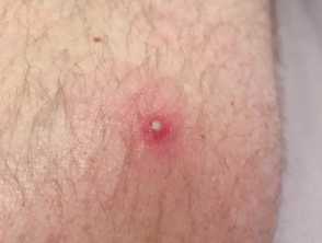
Pathergy is a commonly observed feature in Behçet disease, and refers to the eruption of papulopustules at the site of a needle injury within 24–48 hours. Pathergy is not specific to BD, and can also occur in pyoderma gangrenosum and Sweet syndrome.
Other skin lesions include:
- Acne-like papulopustular and nodular lesions
- Folliculitis
- Pyoderma gangrenosum
- Superficial thrombophlebitis
- Palpable purpura
- Erythema nodosum
- Acute febrile neutrophilic dermatosis (Sweet syndrome).
For more images, see the Behçet disease image page.
Other systemic features
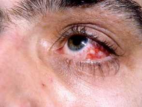
Ophthalmic lesions — seen in 70% of patients, and include:
- Anterior and/or posterior uveitis
- Retinal vasculitis
- Scleritis
- Episcleritis
- Thrombosis of the retinal artery and vein
- Optic neuritis.
Musculoskeletal symptoms include:
- A mono- or oligoarthritis of the medium and large joints
- Fatigue
- Fibromyalgia.
Vascular involvement is present in 25% of patients, resulting in:
- Superficial thrombophlebitis and deep vein thrombosis
- Thrombosis of the vena cava, dural sinus, and Budd-Chiari syndrome
- Arterial manifestations resulting in stenosis, thrombosis, and aneurysm formation.
Involvement of the aorta and pulmonary artery can cause life-threatening occlusion or haemorrhage from aneurysm formation.
Other organ systems which can be affected less commonly include neurological, pulmonary, renal, and cardiac.
How do clinical features vary in differing types of skin?
The features are similar, although Behçet disease is less common in those with skin of colour.
What are the complications of Behçet disease?
Given the multi-system nature of Behçet disease, there are many potential complications.
The most serious complications include:
- Vascular complications, particularly when the aorta and pulmonary arteries are affected — this can be life-threatening
- CNS involvement leading to permanent neurological deficits
- Blindness
- Bowel perforation and peritonitis.
How is Behçet disease diagnosed?
BD can be challenging to diagnose, and several years may elapse before a confident diagnosis can be made, and strict diagnostic criteria fulfilled. It is sometimes useful to classify sufferers as having possible, probable, or definite Behçet disease as new symptoms evolve. There are no definitive confirmatory diagnostic tests.
The International Study Group Criteria are widely used in diagnosis, and criteria include:
- At least three episodes of minor aphthous, major aphthous, or herpetiform oral ulceration over a 12-month period (observed by a doctor or patient) plus at least two of the following:
- Recurrent genital ulceration — aphthous ulceration or scarring observed by a doctor or patient
- Ophthalmic involvement — anterior uveitis, posterior uveitis or cells in vitreous on slit examination or retinal vasculitis observed by an ophthalmologist
- Cutaneous lesions — erythema nodosum observed by a doctor or patient, pseudofolliculitis or papulopustular lesions, or acneiform nodules observed by a doctor
- Positive pathergy test — observed at 24–48 hours.
Common investigations include:
- Full blood count
- Urea and electrolytes
- Syphilis serology
- Antinuclear factor
- Serum iron, folate, zinc and vitamin B12
- Culture/PCR from oral or genital ulcers to exclude HSV infection
- HLA-B51
- Skin biopsy.
Other investigations may be required and depend on the organ system affected.
What is the differential diagnosis for Behçet disease?
This depends on the clinical presentation but other common conditions which cause orogenital aphthous ulcers should be considered.
Differentials include:
- Herpesvirus infections
- Primary and secondary syphilis
- Vitamin B12 deficiency
- Complex aphthosis (recurrent oral and genital ulcers in the absence of other features)
- Inflammatory bowel disease
- Erythema multiforme
- PFAPA syndrome (periodic fever, aphthosis, pharyngitis, adenitis)
- Acute febrile neutrophilic dermatosis
- MAGIC syndrome (mouth and genital ulcers with inflamed cartilage)
- Haploinsufficiency of A20 (HA20)
What is the treatment for Behçet disease?
General principles
Behçet disease treatment should be tailored to each individual patient based on clinical manifestations and extent of disease — an MDT approach is required. Behçet disease typically has a relapsing-remitting course, and treatments should aim to suppress any inflammatory exacerbations and prevent irreversible organ damage, particularly in the initial phase of disease.
Ocular, vascular, and neurological manifestations are often more serious and require more aggressive treatment.
Treatment of mucosal disease
Topical treatments include:
- Corticosteroids applied as creams, gels, or mouth rinses
- Pimecrolimus — as an alternative to topical steroids
- Sucralfate
- Antibiotics such as tetracyclines
- Antiseptics — chlorhexidine mouthwash
- Hydroxypropyl cellulose
- Diclofenac
- Lidocaine.
Systemic treatments for mucosal disease include:
- Colchicine
- Glucocorticoids
- Apremilast
- Azathioprine
- TNF-alpha inhibitors
- Ciclosporin
- Interferon alpha-21 and interferon alpha-2b
- Thalidomide
- Methotrexate
- Secukinumab
- Ustekinumab.
Treatment of skin manifestations
With the exception of erythema nodosum and pyoderma gangrenosum, most other cutaneous lesions respond well to the above treatments. Recalcitrant acneiform lesions may require oral isotretinoin.
Erythema nodosum in Behçet disease:
- Raises the possibility of underlying dermal vasculitis
- Colchicine is generally used first-line, but if this fails, systemic glucocorticoids combined with another immunosuppressive agent may be used.
Evidence of ulceration or detection of medium-vessel vasculitis on skin biopsy is an indication for systemic glucocorticoids with another immunosuppressive agent (typically azathioprine).
Pyoderma gangrenosum in BD should be managed as in other causes of this disease.
Treatment of multi-system Behçet disease
The key management points are:
- Oral corticosteroids: recommended treatment for patients with moderate to severe disease with ocular, vascular, gastrointestinal, or neurologic involvement.
- Azathioprine: as a steroid-sparing agent
- TNF-alpha inhibitors, particularly infliximab for refractory ocular BD
- Cyclophosphamide — reserved for life-threatening complications of BD mainly when there is major vessel or CNS involvement
- Major blood vessel disease, including thrombosis is treated with immunosuppression and not anticoagulation
- CNS involvement is typically more refractory to treatment
- Other agents that can be used include interleukin-1 receptor antagonists (e.g. anakinra), alemtuzumab, tocilizumab, rituximab, and stem cell transplantation.
What is the outcome for Behçet disease?
Behçet disease typically runs a relapsing-remitting course and the prognosis varies depending on the organ systems affected.
Mortality is low but is increased in patients with neurological, vascular, and gastrointestinal involvement. Ophthalmic involvement is a major cause of morbidity.
The prognosis is worse in males.