What are keratinocyte cancers?
A keratinocyte cancer is a malignant neoplasm formed from the epidermal cells that produce keratin (keratinocytes). Keratinocyte cancers are classified as either squamous cell carcinomas (SCC) or basal cell carcinomas (BCC).
Keratinocyte cancers are considered low risk or high risk, depending on patient factors such as age and immune suppression status, and tumour factors including tumour site and histological subtype.
Keratinocyte cancers
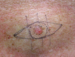
What is a surgical margin?
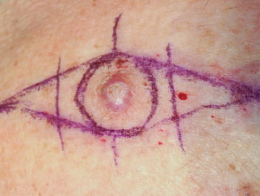
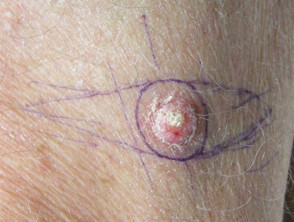
A surgical margin is a clinical measurement made just before a lesion is excised. The surgeon uses their naked eye or magnification (eg, a dermatoscope) to find the visible edge of the tumour, then measures the desired margin of normal skin to remove on either side of the tumour. A surgical marking pen is used to plan the excision site.
As a general guide, adequate surgical margins are 3–4 mm for a BCC and at least 4 mm for a low-risk SCC. There are no published guidelines for a high-risk SCC.
Surgical margins
What are histological margins?
A histological margin is measured by a pathologist. It describes the amount of normal tissue bordering a tumour specimen.
- The lateral and deep margins are examined in several representative histological sections.
- Tumour involving any surgical margin means cancer cells are seen at the edge of the specimen.
- The number of sections evaluated varies and may represent as little as 1% of the tumour border.
Why is the histological margin important?
Histological margins may be adequate, narrow, or inadequate.
- Narrow or inadequate margins may require close monitoring, further surgery, or adjunctive therapy (eg, radiotherapy).
- Histological margins may have prognostic value, including the likelihood of recurrence and metastasis.
What is an adequate histological margin?
An adequate histological margin is the acceptable distance of tumour cells from the histological margin, according to expert consensus and published research.
The desirable margin depends on the type of tumour and the technique used to remove it. Clinicopathological correlation and additional sections may be required, particularly when clinical margins have been difficult to assess or compromised by the anatomical location of the tumour (eg, a lesion close to the eye).
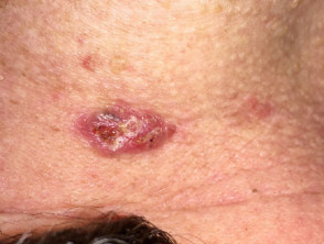
What are the high-risk features of keratinocyte cancers?
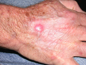
Basal cell carcinoma
High-risk BCC may be defined by any of the following features:
- Size ≥ 20 mm on the trunk or extremities or ≥ 10 mm on cheeks, forehead, scalp, neck, and pretibial sites
- Any lesion on the central face, eyelids, eyebrows, periorbital area, nose, lips, chin, mandible, postauricular area, genitalia, hands, or feet
- Any aggressive histological subtype — morphoeaform, sclerosing, mixed infiltrative, micronodular, or basosquamous BCC
- Any tumour with perineural invasion
- Any recurrent tumour
- Any tumour in an immunocompromised patient
- Any tumour in an area of previous radiotherapy
- Any tumour with poorly defined borders [4].
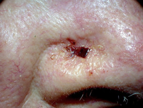
High-risk basal cell carcinoma
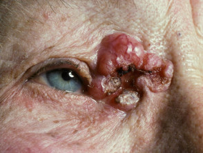
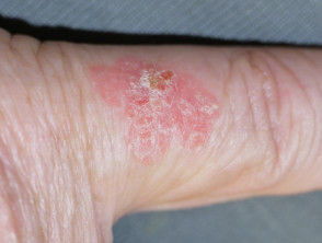
Squamous cell carcinoma
High-risk SCC may be defined by any of the following features:
- Size ≥ 20 mm on the trunk or extremities or size ≥ 10 mm on the cheeks, forehead, scalp, neck, and pretibial sites
- Any lesion on the central face, eyelids, eyebrows, periorbital area, nose, lips, chin, mandible, ear, postauricular area, genitalia, hands, or feet
- Any poorly differentiated tumour
- Any tumour ≥ 4 mm in thickness on biopsy
- Any tumour with perineural, lymphatic, or vascular invasion
- Any tumour in an immunocompromised patient
- Any recurrent tumour [5].
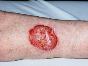
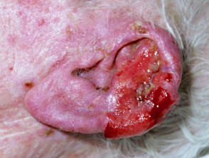
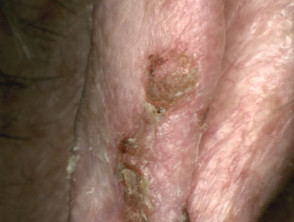
High-risk squamous cell carcinoma
Perineural invasion
Perineural invasion may be seen on histology in up to 5% of keratinocyte cancers. Depending on the size of the nerves involved, it may pose a high-risk for local recurrence and, in SCC, for metastasis [6].
Tumours with perineural invasion should be referred to a specialist. Management may include:
- Magnetic resonance imaging (MRI)
- Re-excision with wide margins
- Postoperative radiotherapy.
What is the treatment if margins are inadequate?
Basal cell carcinoma
If the histological margins are involved or inadequate, most low-risk BCCs should undergo re-excision to ensure clear margins. In select cases, radiotherapy may be a reasonable alternative. Persistent superficial BCC may occasionally be treated topically (eg, imiquimod cream).
Inadequately excised high-risk lesions should be referred to a specialist, depending on the clinician’s skill and tumour factors. To reduce unnecessary removal of healthy tissue, Mohs micrographic surgery may be considered, especially for lesions in high-risk areas.
Squamous cell carcinoma
Histological margins of less than 1 mm should lead to re-excision of an SCC. High-risk SCCs, especially on the head and neck, require referral to a specialist with expertise in this field.
How common is a recurrence of keratinocyte cancer?
Recurrence of a keratinocyte cancer depends on the type and risk category of the lesion, the type of treatment, the histological margins, and the clinician’s expertise.
For low-risk BCCs treated by surgical excision:
- In one study, recurrence rates with 5, 4, 3, and 2-mm surgical margins were 0.4%, 1.6%, 2.6%, and 4% respectively, and 27% recurred if the pathologic margin was involved [7]
- In another study, the recurrence rate was 38% if histological margins were involved, 12% if histological margins were < 0.5 mm, and 1.2 % for margins of 0.5 mm [8].
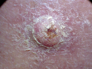
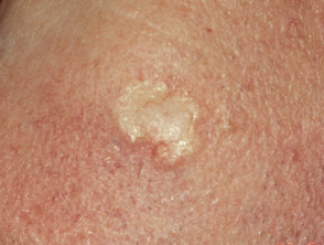
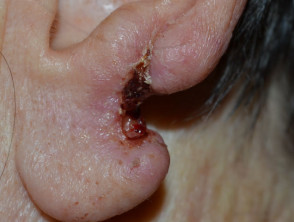
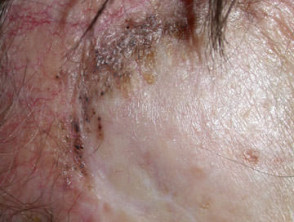
Recurrent basal cell carcinoma
What are the complications due to recurrent keratinocyte cancers?
Basal cell carcinoma
BCCs rarely metastasise but can be locally invasive and cause local tissue destruction. Recurrent BCCs usually require more aggressive treatment resulting in greater tissue loss and surgical complications.
Squamous cell carcinoma
Development of local recurrence of SCC is associated with further local recurrence in about 25% of lesions and subsequent metastasis to regional nodes in up to 30%. About a third of patients who develop regional metastasis will die from their SCC [9].
What is the treatment for recurrent keratinocyte cancers?
Basal cell carcinoma
Recurrent BCC is treated by resection of the scar, the recurrent tumour, and a 3–4 mm margin of surrounding skin [10,11]. Specialist referral should be considered, especially if the tumour edges are hard to detect. Mohs micrographic surgery may be indicated.
Squamous cell carcinoma
Specialist referral is recommended for recurrent SCC due to the risk of developing further recurrence and metastasis.
What is the follow-up for recurrent keratinocyte cancer?
Follow-up requires inspection of the treated area using dermoscopy and palpation of adjacent structures [12].
- 50% of BCC recurrences occur within the first year and 80% within 5 years [13].
- 70–80% of SCC recurrences occur in the first two years and ~95% within 5 years [9].
An annual skin check is recommended for all patients with a history of skin cancer.
- No additional follow-up is required after the complete excision of low-risk SCC and BCC [2].
- Follow-up for a BCC with high-risk features should be every 6 months for the first year than annually [4].
- More intense follow-up is recommended after non-surgical treatments have been used (eg, for superficial BCC) due to a higher recurrence rate, reviewing after 3 months, then reviewing every 6–12 months for 3 years [2].
- Follow-up for SCC with high-risk features is recommended after 3 months then every 6 months; always examine the draining lymph node basin [2].