DermNet provides Google Translate, a free machine translation service. Note that this may not provide an exact translation in all languages

Translate

Seborrhoeic keratosis

Author: Dr Amanda Oakley, Dermatologist, Hamilton, New Zealand, 1997. Updated, January 2016.


toc-icon

What is a seborrhoeic keratosis?

Seborrhoeic keratosis is a harmless warty spot that appears during adult life as a common sign of skin ageing. Some people have hundreds of them.

Seborrhoeic keratosis (American spelling - seborrheic keratosis) is also called SK, basal cell papilloma, senile wart, brown wart, wisdom wart, or barnacle.  The descriptive term, benign keratosis, is a broader term that is used to include the following related scaly skin lesions:

  • Seborrhoeic keratosis 
  • Solar lentigo (which can be difficult to distinguish from a flat seborrhoeic keratosis)
  • Lichen planus-like keratosis (which arises from a seborrhoeic keratosis or a solar lentigo).

Seborrhoeic keratoses

Who gets seborrhoeic keratoses?

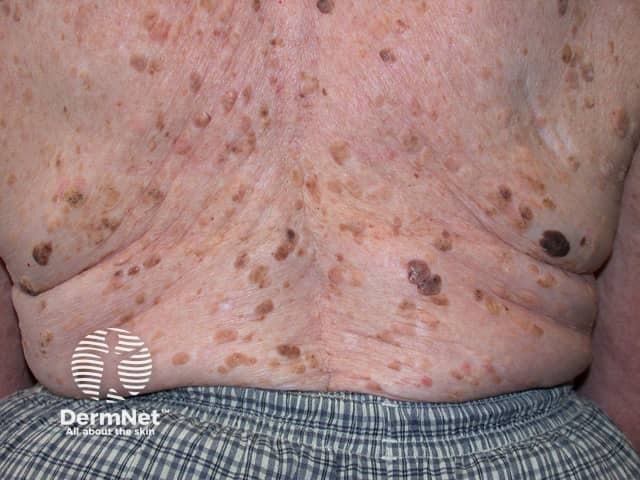
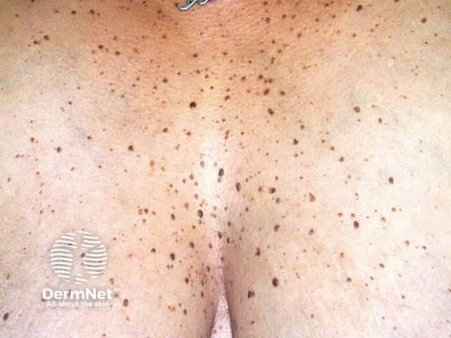
Seborrhoeic keratoses are extremely common. It has been estimated that over 90% of adults over the age of 60 years have one or more of them. They occur in males and females of all races, typically beginning to erupt in the 30s or 40s. They are uncommon under the age of 20 years.

What causes seborrhoeic keratoses?

The precise cause of seborrhoeic keratoses is not known.

The name is misleading, because they are not limited to a seborrhoeic distribution (scalp, mid-face, chest, upper back) as in seborrhoeic dermatitis, nor are they formed from sebaceous glands, as is the case with sebaceous hyperplasia, nor are they associated with sebum — which is greasy.

Seborrhoeic keratoses are considered degenerative in nature. As time goes by, seborrhoeic keratoses become more numerous. Some people inherit a tendency to develop a very large number of them. Researchers have noted:

  • Eruptive seborrhoeic keratoses can follow sunburn or dermatitis.
  • Skin friction may be the reason they appear in body folds.
  • Viral cause (eg human papillomavirus) seems unlikely.
  • Stable and clonal mutations or activation of FRFR3, PIK3CA, RAS, AKT1 and EGFR genes are found in seborrhoeic keratoses.
  • Seborrhoeic keratosis can arise from solar lentigo.
  • FRFR3 mutations also arise in solar lentigines. These mutations are associated with increased age and location on the head and neck, suggesting a role of ultraviolet radiation in these lesions.
  • Seborrhoeic keratoses do not harbour tumour suppressor gene mutations.
  • Epidermal growth factor receptor inhibitors (used to treat cancer) often result in an increase in verrucal (warty) keratoses.

What are the clinical features of seborrhoeic keratoses?

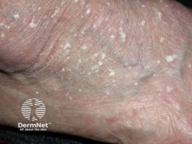
Seborrhoeic keratoses can arise on any area of skin, covered or uncovered, with the exception of palms and soles. They do not arise from mucous membranes.

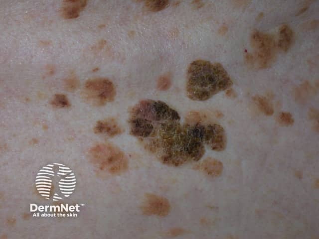
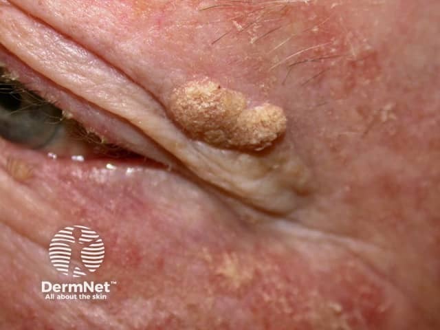
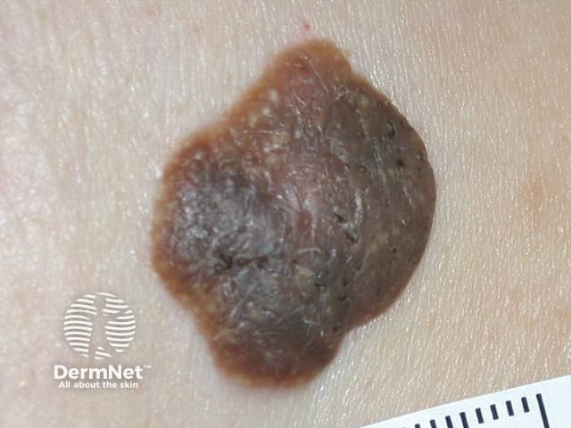
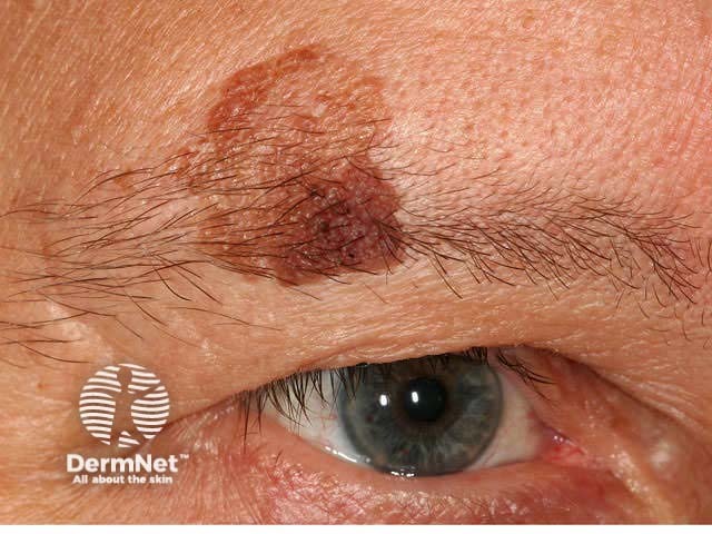
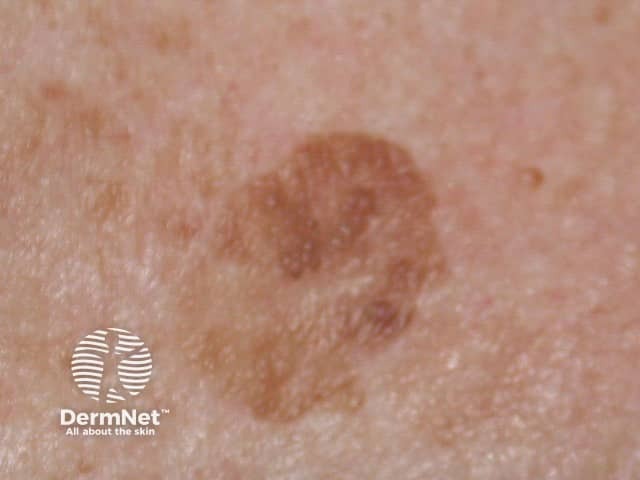
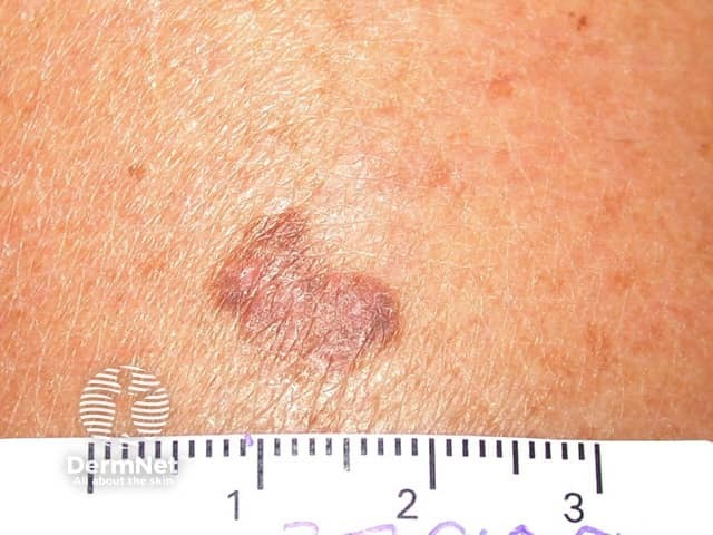
Seborrhoeic keratoses have a highly variable appearance.

  • Flat or raised papule or plaque
  • 1 mm to several cm in diameter
  • Skin coloured, yellow, grey, light brown, dark brown, black or mixed colours
  • Smooth, waxy or warty surface
  • Solitary or grouped in certain areas, such as within the scalp, under the breasts, over the spine or in the groin

They appear to stick on to the skin surface like barnacles.

Seborrhoeic keratoses

Variants of seborrhoeic keratoses

Variants of seborrhoeic keratoses include:

  • Solar lentigo: flat circumscribed pigmented patches in sun-exposed sites
  • Dermatosis papulosa nigra: small, pedunculated and heavily pigmented seborrhoeic keratoses on head and neck of darker-skinned individuals
  • Stucco keratoses: grey, white or yellow papules on the lower extremities
  • Inverted follicular keratosis
  • Large cell acanthoma
  • Lichenoid keratosis: an inflammatory phase preceding involution of some seborrhoeic keratoses and solar lentigines.

Benign keratoses

Complications of seborrhoeic keratoses

Seborrhoeic keratoses are not premalignant tumours. However:

  • Skin cancers are sometimes difficult to tell apart from seborrhoeic keratoses.
  • Skin cancer may by chance arise within or collide with a seborrhoeic keratosis.

Very rarely, eruptive seborrhoeic keratoses may denote an underlying internal malignancy, most often gastric adenocarcinoma. The paraneoplastic syndrome is known as the sign of Leser-Trélat. Eruptive seborrhoeic keratoses that are not associated with cancer are sometimes described as having pseudo-sign of Leser-Trélat.

Eruptive and irritated seborrhoeic keratoses may also arise as an adverse reaction to a medication, such as adalimumab, vemurafenib, dabrafenib, 5-fluorouracil and many chemotherapy drugs.

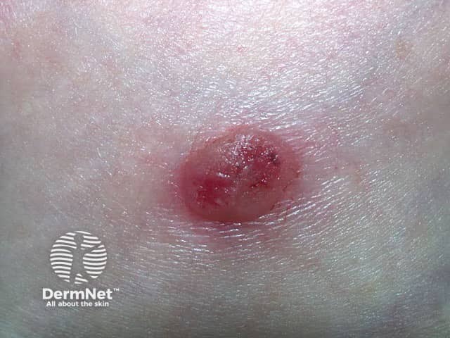
An irritated seborrhoeic keratosis is an inflamed, red and crusted lesion. It may give rise to eczematous dermatitis around the seborrhoeic keratosis. Dermatitis may also trigger new seborrhoeic keratoses to appear.

How is a seborrhoeic keratosis diagnosed?

The diagnosis of seborrhoeic keratosis is often easy.

  • A stuck-on, well-demarcated warty plaque
  • Other similar lesions

Sometimes, seborrhoeic keratosis may resemble skin cancer, such as basal cell carcinoma, squamous cell carcinoma or melanoma.

Dermoscopy often shows a disordered structure in a seborrhoeic keratosis, as is also true for skin cancer. There are diagnostic dermatoscopic clues to seborrhoeic keratosis, such as multiple orange or brown clods (due to keratin in skin surface crevices), white milia-like clods, and curved thick ridges and furrows forming a brain-like or cerebriform pattern.

If doubt remains, a seborrhoeic keratosis may undergo partial shave or punch biopsy or diagnostic excision. [see Seborrhoeic keratosis pathology]

The dominant histopathological features of seborrhoeic keratosis may be described as:

  • Melanoacanthoma (deeply pigmented)
  • Acanthotic
  • Hyperkeratotic or papillomatous
  • Adenoid or reticulated
  • Clonal or nested
  • Adamantinoid or mucinous
  • Desmoplastic
  • Irritated.

What is the treatment for seborrhoeic keratoses?

An individual seborrhoeic keratosis can easily be removed if desired. Reasons for removal may be that it is unsightly, itchy, or catches on clothing.

Methods used to remove seborrhoeic keratoses include:

All methods have disadvantages. Treatment-induced loss of pigmentation is a particular issue for dark-skinned patients. There is no easy way to remove multiple lesions on a single occasion.

How can seborrhoeic keratoses be prevented?

How to prevent seborrhoeic keratoses is unknown.

What is the outlook for seborrhoeic keratoses?

Seborrhoeic keratoses tend to persist. From time to time, individual or multiple lesions may remit spontaneously or via the lichenoid keratosis mechanism.

Those associated with dermatitis may regress after it has been controlled.


References

  • Jackson JM, Alexis A, Berman B, Berson DS, Taylor S, Weiss JS. Current Understanding of Seborrheic Keratosis: Prevalence, Etiology, Clinical Presentation, Diagnosis, and Management. J Drugs Dermatol. 2015 Oct 1;14(10):1119–25. PubMed
  • Hafner C, Hartmann A, van Oers JM, Stoehr R, Zwarthoff EC, Hofstaedter F, Landthaler M, Vogt T. FGFR3 mutations in seborrheic keratoses are already present in flat lesions and associated with age and localization. Mod Pathol. 2007 Aug;20(8):895–903. Epub 2007 Jun 22. PubMed
  • Hafner C, Hafner H, Groesser L. [Genetic basis of seborrheic keratosis and epidermal nevi]. Pathologe. 2014 Sep;35(5):413–23. doi: 10.1007/s00292-014-1928-9. Review. German. PubMed
  • Hida Y, Kubo Y, Arase S. Activation of fibroblast growth factor receptor 3 and oncogene-induced senescence in skin tumours. Br J Dermatol. 2009 Jun;160(6):1258–63. doi: 10.1111/j.1365-2133.2009.09068.x. Epub 2009 Mar 9. PubMed
  • Husain Z, Ho JK, Hantash BM. Sign and pseudo-sign of Leser-Trélat: case reports and a review of the literature. J Drugs Dermatol. 2013 May;12(5):e79–87. Review. PubMed

On DermNet

Other websites

Books about skin diseases

Text: Miiskin


Related information

Sign up to the newsletter