What is a subepidermal calcified nodule?
Subepidermal calcified nodules are deposits of insoluble calcium or phosphorus in the skin. Also known as Winer’s nodular calcinosis or solitary congenital nodular calcification, they are a subtype of calcinosis cutis.
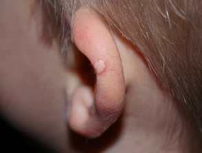
A subepidermal calcified nodule on the ear - note the typical chalky appearence
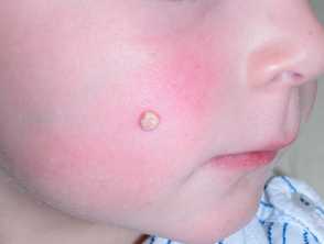
A subepidermal calcified nodule on the cheek - cheeks and ears are the most frequent location
A subepidermal calcified nodule on the cheek
A chalky-white gritty nodule on the ear due to a subepidermal calcified nodule
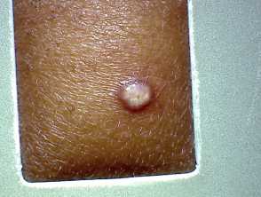
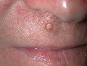
A subepidermal calcified nodule on the lip of an adult - they are more common in children
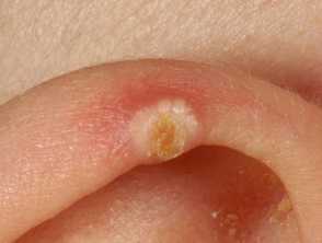
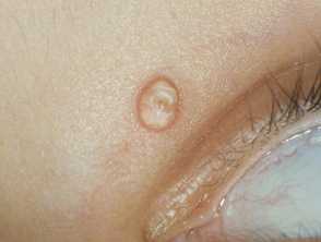
A subepidermal calcified nodule on the lid
Who gets subepidermal calcified nodules?
Subepidermal calcified nodules are typically found in children, although almost 30% of cases have been reported in adults. They are relatively rare with only a few hundred cases reported. They are twice as common in men as in women.
What causes subepidermal calcified nodules?
As a type of idiopathic calcinosis cutis, subepidermal calcified nodules are of unclear aetiology. While suggested to be a form of dystrophic calcification, controversy remains over the exact mechanism.
They occur in supposedly undamaged skin (although some have suggested that unappreciated trauma may be responsible), in patients with normal serum calcium, phosphate, and parathyroid hormone levels.
What are the clinical features of subepidermal calcified nodules?
- Solitary nodules in 80% of cases, but can be multiple.
- Painless, firm nodules that are mobile from underlying structures.
- Typically asymptomatic, but sometimes bleed.
- Usually present as yellow-white papules and may be smooth or verrucous.
- Preponderance for the head and neck and often affect the eyelid.
Difficult to differentiate from other cutaneous nodules via general examination, dermoscopy reveals yellow-white clods, surrounded by linear vessels.
How do clinical features vary in differing types of skin?
A firm, mobile, asymptomatic nodule is characteristic in case reports of subepidermal calcified nodules in different skin types, with no specific differences observed.
What are the complications of subepidermal calcified nodules?
Subepidermal calcified nodules are benign lesions, although they may cause both cosmetic concerns or local irritation and pain.
How is a subepidermal calcified nodule diagnosed?
As clinical examination often leads to misdiagnosis, histology may be the only way of confirming diagnosis.
Histologically, subepidermal calcified nodules show:
- In younger patients — a dermis with multiple small, calcified globules surrounded by giant cells and chronic lymphohistiocytic inflammation
- In older patients — typically a large singular calcium deposit with a fibrous capsule, with no chronic inflammatory process.
Recently, reflectance confocal microscopy (RCM) has also been suggested as a potential diagnostic tool.
What is the differential diagnosis for a subepidermal calcified nodule?
What is the treatment for a subepidermal calcified nodule?
Lesions can be managed expectantly if asymptomatic, or can be surgically excised. Excision usually results in complete removal without recurrence.
Curettage, laser, salicylic acid, and intralesional triamcinolone have all been described.
What is the outcome for subepidermal calcified nodules?
Subepidermal calcified nodules are benign. If elective excision is chosen, almost always for cosmetic reasons or pain, outcomes are excellent and recurrence has not been described.