DermNet provides Google Translate, a free machine translation service. Note that this may not provide an exact translation in all languages

Translate

Trichilemmal cyst pathology

Author: Dr Ben Tallon, Dermatologist/Dermatopathologist, Tauranga, New Zealand, 2012.


toc-icon
Table of contents
arrow-right-small

Trichilemmal cyst is also known as pilar cyst. It is seen on the scalp and multiple cysts are common.

Histology of trichilemmal cyst

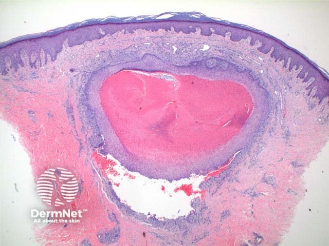
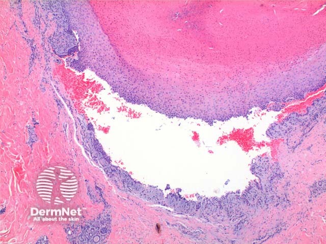
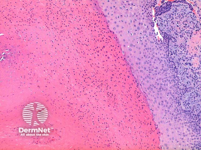
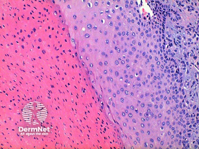
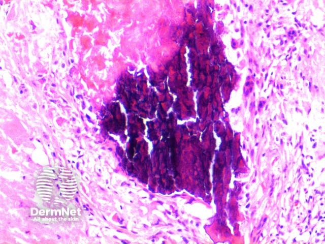
Scanning power view of trichilemmal cyst shows a epithelial lined cyst filled with brightly eosinophilic keratinaceous debris (Figure 1). Focal rupture of the cyst may occur with an associated giant cell reaction (Figure 2). Closer inspection of the cyst wall identifies trichilemmal differentiation (Figures 3 and 4) as occurs in the outer root sheath of the hair follicle. This is seen as maturation of squamous epithelium with lack of a granular layer. The eosinophilic keratin centrally is densely packed frequently displaying cholesterol clefts. Focal calcification is seen in around 25% of cases (Figure 5).

Trichilemmal cyst pathology

Histological variants of trichilemmal cyst

  • Proliferating trichilemmal cyst: In this variant squamous proliferation can be seen arising from the cyst wall.
  • Malignant proliferating trichilemmal tumour: This tumour arises out of a pre-existing trichilemmal cyst. Clear transition is evident into an area of eccentric asymmetrical growth with malignant cytology

Differential diagnosis

Epidermal inclusion cyst: This cyst retains a granular layer within the maturing epithelial cyst wall.

Vellus hair cyst: The cyst contents contain multiple vellus hairs seen as small non pigmented hair shafts.


References

  • Skin Pathology (3rd edition, 2002). Weedon D
  • Pathology of the Skin (3rd edition, 2005). McKee PH, J. Calonje JE, Granter SR

On DermNet

Books about skin diseases


Related information

Sign up to the newsletter