The deposition of calcium in the skin, subcutaneous tissue, muscles and visceral organs is known as calcinosis. This condition commonly occurs in the skin, where it is known as calcinosis cutis or cutaneous calcification.
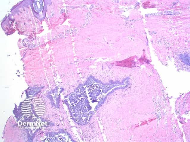
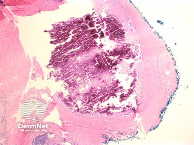
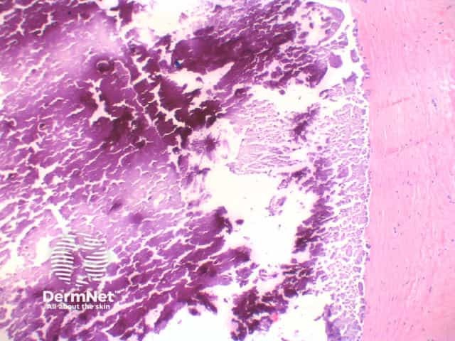
There are irregular deposits of intensely basophilic acellular material in the dermis and subcutaneous tissue (figure 1). The basophilia is so strong that the appearance is of a deep purple (figures 2, 3). The deposits are typically well circumscribed, with a thin rim of eosinophilic hyalinisation and frequently a host giant cell reaction.
Artefactual lines form in the calcification and surrounding tissue due to shatter with cutting (figure 1).
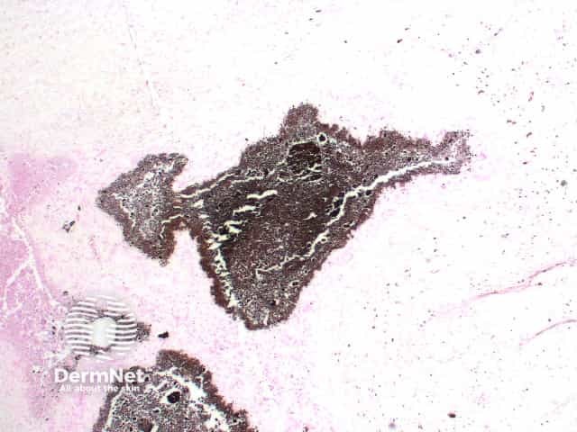
The Von Kossa silver stain highlights the calcium salts (black). Figure 4.
Osteoma cutis: The deposits here are eosinophilic with visible osteocytes sitting within small lacunae.
Gouty tophi: The crystal deposits form washed out pale areas surrounded by a dense inflammatory cell infiltrate containing multinucleated giant cells. Needle like spaces can be seen in a radial array.

