DermNet provides Google Translate, a free machine translation service. Note that this may not provide an exact translation in all languages

Translate

Dystrophic epidermolysis bullosa

Author: Jane Widdowson et al, DEBRA New Zealand, February 2016. Minor update October 2023.


toc-icon

What is epidermolysis bullosa?

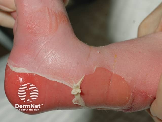
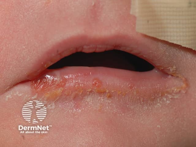
Epidermolysis bullosa (EB) is a group of inherited diseases that are characterised by blistering lesions on the skin and mucous membranes. These may occur anywhere on the body but most commonly appear at sites of friction and minor trauma such as the feet and hands. In some subtypes, blisters may also occur on internal organs, such as the oesophagus, stomach and respiratory tract, without any apparent friction.

What is dystrophic epidermolysis bullosa?

Dystrophic epidermolysis bullosa (DEB) is characterised by the site of blister formation in the lamina densa within the basement membrane zone and the upper dermis. It causes generalised blistering of the skin and internal mucous membranes and leads to scar formation.

Who gets dystrophic epidermolysis bullosa?

Dystrophic epidermolysis bullosa is a rare inherited disease. There are two main subtypes: autosomal dominant dystrophic epidermolysis bullosa (DDEB) and autosomal recessive dystrophic epidermolysis bullosa (RDEB). The latter is the more severe form.

What is the cause of dystrophic epidermolysis bullosa?

Dominant dystrophic epidermolysis bullosa is caused by heterozygous mutation in the type VII collagen gene (COL7A1; 120120) on chromosome 3p21.

Recessive dystrophic epidermolysis bullosa is due to homozygous or compound heterozygous mutation in the gene encoding type VII collagen (COL7A1; 120120) on chromosome 3p21.

What are the clinical features of dystrophic epidermolysis bullosa?

DEB Subtypes Features
Dominant generalised EB (DEB)
  • Generalised blistering present at birth
  • Blistering becomes localised to hands, feet, elbow or knees as the child grows older and in response to friction
  • Small white spots called milia are often present at healed but scarred sites
  • Bart syndrome: aplasia cutis, lesions in the mouth, and abnormal nails due to abnormal type 7 collagen in anchoring fibrils
  • May also get blistering of the oesophagus
Generalised severe recessive EB (RDEB)
Previously known as Hallopeau-Siemens; and;
Generalised intermediate RDEB (previously Non-Hallopeau-Siemens)
  • May present with severe blistering (generalised severe RDEB) or mild disease (generalised intermediate RDEB)
  • Generalised severe blistering is more common and involves large areas of skin and mucous membranes
  • Blisters heal but with scarring and deformity causing limited movement as fingers and toes may be fused together (mitten hands)
  • Complications such as infection, malnutrition and dehydration may cause death in infancy
  • Those that survive are at great risk of developing potentially dangerous squamous cell carcinoma (SCC) within chronic EB wounds. SCC look and behave differently in EB from in unaffected individuals. They are found on covered sites and grow rapidly. They present as ulcerated or keratotic nodules and plaques.

Recessive dystrophic epidermolysis bullosa

How is dystrophic epidermolysis bullosa diagnosed?

In the dominant subtypes of epidermolysis bullosa, where an informative family tree is known, it is often acceptable for clinical diagnosis to be made by a specialist dermatologist based on the presenting signs.

  • Diagnostic tests are also available in some countries and include skin biopsy of a newly induced blister which undergoes immunofluorescence antigen mapping (IFM) and/or transmission electron microscopy (EM). Mutational analysis (blood testing of genes), although not currently considered the first-line diagnostic test, is also available in some countries.

Squamous cell carcinoma in dystrophic epidermolysis bullosa is diagnosed by its clinical appearance and supported by biopsy.

What is the treatment of dystrophic epidermolysis bullosa?

See treatment of epidermolysis bullosa – general.

  • Puritus (itching) may be a problem for patients with dystrophic epidermolysis bullosa — strategies such as moisturising and avoiding hot environments can help.
  • Oleogel-S10, containing birch triterpenes or birch bark extract, has been shown to accelerate the healing of chronic wounds in junctional and dystrophic epidermolysis bullosa.
  • In dominant dystrophic epidermolysis bullosa (DDEB), the focus is on blister prevention and management. This refers to the skin, but may also include eating soft foods to reduce oesophageal blisters.
  • In severe recessive dystrophic epidermolysis bullosa (RDEB), high attention to both the skin and mucous membranes is necessary. Management of pain, pruritus, infection, scarring and deformities, malnutrition and anaemia all play a major part in the day to day treatment of RDEB.
  • Squamous cell carcinomas are treated surgically. This should be done early, as the tumours are aggressive and have often metastasised by the time of diagnosis. Amputation may be required.

What is the outcome for patients with dystrophic epidermolysis bullosa?

Life expectancy is unaffected in dominant dystrophic epidermolysis bullosa. In recessive dystrophic epidermolysis bullosa, life expectancy has significantly improved due to appropriate management and interventions related to complications of the disease including the early detection and treatment of squamous cell carcinoma (SCC).


References

On DermNet

Other websites

Books about skin diseases


Related information

Sign up to the newsletter