What is indirect immunofluorescence?
Indirect immunofluorescence, or secondary immunofluorescence, is a technique used in laboratories to detect circulating autoantibodies in patient serum. It is used to diagnose autoimmune blistering diseases.
- Unlabelled primary antibodies (used as a marker for cancer, diabetes and Alzheimer disease) from the patient serum bind to the target molecule in pre-prepared tissue samples.
- Secondary antibodies (used as a marker for HIV), conjugated with a fluorescent dye such as fluorescein isothiocyanate (FITC), bind to the primary antibody.
- When exposed to light, the fluorescent dye is excited and emits a wavelength that can be seen with a fluorescent microscope.
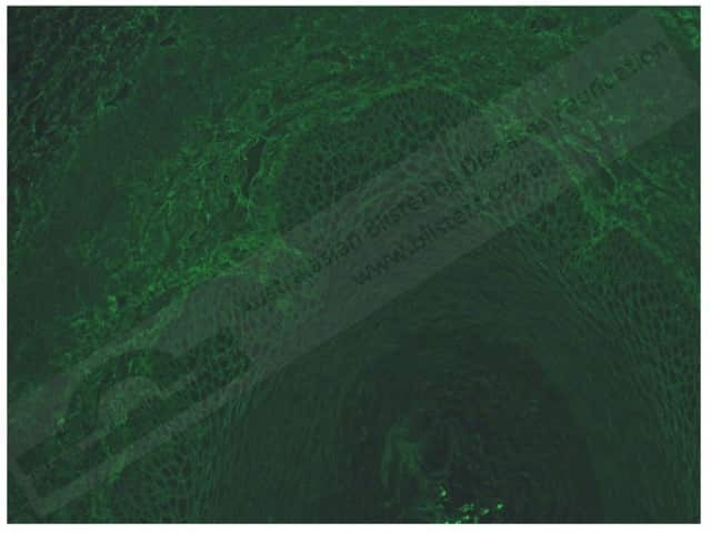
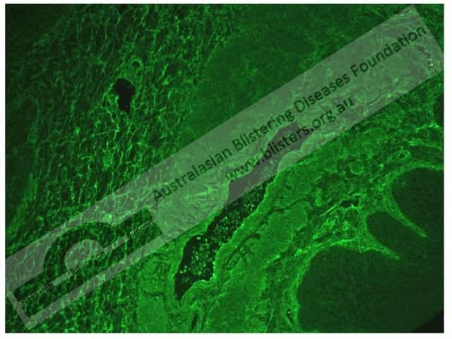
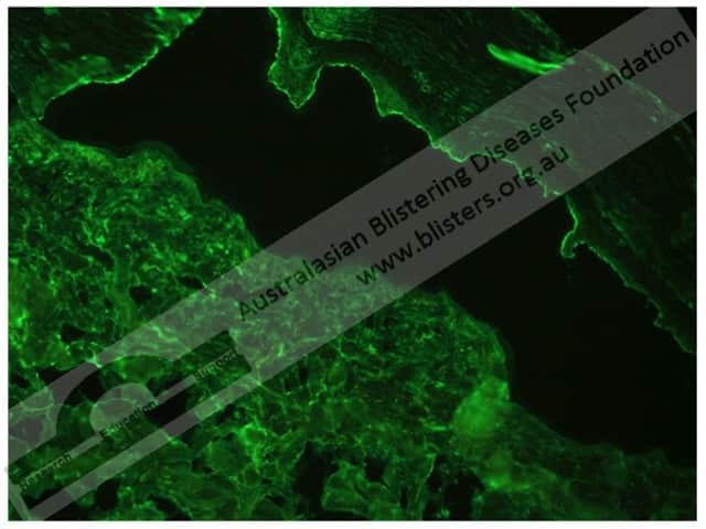
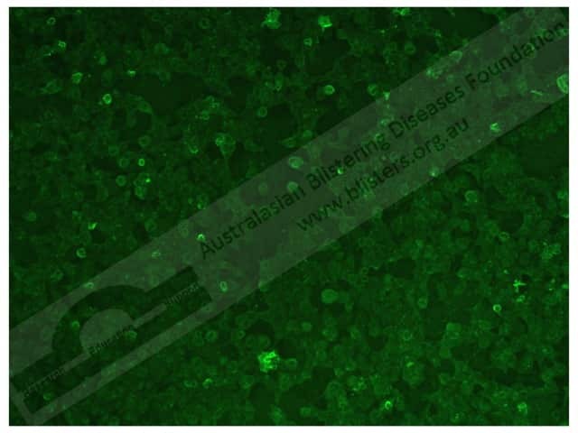
The tissue substrates used (whether monkey oesophagus or human skin) will depend on the suspected disease. Different staining patterns will help diagnose an autoimmune bullous disease.
Which blistering skin conditions does an indirect immunofluorescence test for?
Indirect immunofluorescence is used to diagnose the following autoimmune bullous diseases:
- Pemphigus vulgaris
- Pemphigus foliaceus
- Paraneoplastic pemphigus
- Immunoglobulin (Ig)A pemphigus
- Linear IgA bullous dermatosis and chronic bullous disease of childhood
- Dermatitis herpetiformis
- Bullous pemphigoid
- Pemphigoid gestationis
- Mucous membrane pemphigoid
- Epidermolysis bullosa acquisita
- Bullous systemic lupus erythematosus.
The laboratory method
Preparing patient serum for testing
- 5–10 mL of blood is drawn and collected in a tube.
- The blood is allowed to clot.
- The blood sample is then centrifuged to separate the serum from the clot.
- The serum is divided into parts and frozen until testing.
The indirect immunofluorescence test
- The tissue is cut into 4–6 micron thick slices for the tissue substrate slides (this can be done in a laboratory, or the slides can be obtained commercially).
- Several slides are prepared with different tissue substrates.
- The tissue sections are incubated with patient serum for 30 minutes and doubling dilution is performed if titres are required.
- The slides are washed to remove any unbound primary antibodies.
- The slides are incubated for another 30 minutes with secondary antibodies containing a fluorescent dye.
- Each slide is mounted under a coverslip.
- Each slide is examined using fluorescence microscopy.
Interpreting the results of indirect immunofluorescence
The immunofluorescent slides are examined to determine the presence of autoantibodies via the patterns of immune deposition. The results are subjective and indirect immunofluorescence cannot be used reliably to monitor disease severity and its treatment.
The different staining patterns seen with indirect immunofluorescence include the:
- Intercellular space (ICS) staining pattern
- Basement membrane zone (BMZ) staining pattern.
The sensitivity of a diagnostic tool refers to its ability to accurately identify those with the disease (ie, producing a true-positive result). The specificity of a diagnostic tool refers to its ability to accurately identify those without the disease (ie, producing a true-negative result).
Occasionally a test can also produce false results. Hence:
- The result may be negative even if the patient has the disease (ie, producing a false-negative result).
- The result may be positive in a patient without the disease (ie, producing a false-positive result).
Immunofluorescent patterns of skin disease
The immunofluorescent patterns seen in the following skin diseases are described below.
Pemphigus vulgaris
In pemphigus vulgaris, indirect immunofluorescence shows:
- An ICS staining pattern with IgG or C3 deposits
- A sensitivity of 81–87% [4,7]
- A specificity of 81% [7].
Pemphigus foliaceous
In pemphigus foliaceus, indirect immunofluorescence shows:
- ICS pattern IgG or C3
- 60-86% [4,7].
Paraneoplastic pemphigus
In pemphigus, indirect immunofluorescence shows:
- C3 and IgG antibodies
- A sensitivity of 80% [3]
- A specificity of 83% [3].
Bullous pemphigoid
In bullous pemphigoid, indirect immunofluorescence shows:
- A linear BMZ staining pattern with IgG and C3 deposits
- A sensitivity of 70% [3].
Mucous membrane pemphigoid
In mucous membrane pemphigoid, indirect immunofluorescence shows:
- A BMZ staining pattern with IgG or IgA antibodies
- A sensitivity of 20% [3].
Pemphigoid gestationis
In pemphigoid gestationis, indirect immunofluorescence shows:
- C3 deposits
- A sensitivity of 93% [3].
Epidermolysis bullosa acquisita
In epidermolysis bullosa acquisita, indirect immunofluorescence shows:
- A linear BMZ staining pattern with IgG antibodies
- A sensitivity of 50% [3].
Dermatitis herpetiformis
In dermatitis herpetiformis, indirect immunofluorescence shows:
- IgA anti-endomysial antibodies
- A sensitivity of 80% [3].
Linear IgA bullous dermatosis and chronic bullous disease of childhood
In linear IgA bullous dermatosis and chronic bullous disease of childhood, indirect immunofluorescence shows:
- BMZ with IgA antibodies
- A sensitivity of 50% [3].
Bullous lupus erythematosus
In bullous systemic lupus erythematosus, indirect immunofluorescence shows a linear BMZ pattern with IgG antibodies.