DermNet provides Google Translate, a free machine translation service. Note that this may not provide an exact translation in all languages

Translate

Paraneoplastic pemphigus

August 2022

Author: Dr Shilpa Manupati and Dr Farazmand Paymaneh, Speciality Doctors in Dermatology, U.K. (2022)
Previous contributors: Vanessa Ngan (2003) 
Reviewing dermatologist: Dr Ian Coulson

Edited by the DermNet content department


toc-icon

What is paraneoplastic pemphigus?

Paraneoplastic pemphigus (PNP) is a rare, autoimmune, mucocutaneous, blistering disease that is almost always associated with a confirmed or occult neoplasm.

Paraneoplastic autoimmune multiorgan syndrome (PAMS) is another proposed name for PNP due to potential autoimmune reactions also appearing in internal organs, such as bronchiolitis obliterans in the lung.

Paraneoplastic pemphigus

For more images click here

Who gets paraneoplastic pemphigus?

PNP is a rare autoimmune bullous disease variant; it is the least common but most serious form of pemphigus, accounting for 3–5% of all cases. Though PNP most commonly develops in adults between 45–70 years of age with equal male and female sex predilection, it is also seen in children.

Almost all cases of PNP are associated with an underlying cancerous (malignant) process:

  • Approximately 5% of patients with internal malignancy may develop PNP
  • Lymphoproliferative neoplasms are the most common underlying disease (~84% of PNP cases): specifically, non-Hodgkin lymphoma is the most common type of underlying cancer in adults, followed by chronic lymphocytic leukaemia (CLL)
  • PNP is often the first presenting feature of Castleman disease (giant cell or angiofollicular lymph node hyperplasia) in children and adolescents.

What causes paraneoplastic pemphigus?

The cause of PNP is not completely understood, but it appears that underlying malignancy (most commonly a lymphoproliferative neoplasm) stimulates an autoimmune response:

  • Autoreactive T-cell generation by neoplasms can induce both humoral and cell-mediated immunity
  • Components of desmosomes (which interconnect the keratinocytes in the epidermis) appear to be the primary autoantigenic targets in PNP (e.g. envoplakin, periplakin, epiplakin, desmoplakin, desmoglein, and desmocollin)
  • Autoantibodies against components of hemidesmosomes (which anchor the epidermis to the dermis), such as bullous pemphigoid (BP) autoantigens BP230 and BP180 and plectin, may also be seen in PNP
  • Humoral immunity, through IgG1 autoantibodies against desmosome and hemidesmosome components, can cause acantholysis, resulting in intraepidermal  blistering and bronchiolitis obliterans 
  • Cell-mediated immunity, through interferon-γ (IFN-γ) and cytotoxic molecules like granzyme and perforin, can also contribute to bronchiolitis obliterans, as well as lichenoid dermatitis.

Genetic factors also play a role in the autoimmune response:

  • People with certain gene variants (alleles), such as the HLA Class II DRB1*03 allele and the HLA-Cw*14 allele (more common in those of Caucasian and Chinese ethnicity respectively), have an increased risk of developing PNP. 

What are the clinical features of paraneoplastic pemphigus?

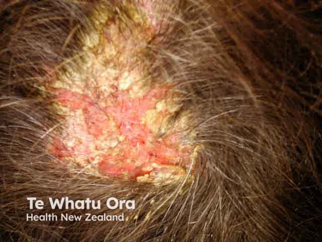
Clinical features may include:

  • Extensive refractory mucostomatitis (often the only presenting feature of PNP), with painful mucous membrane erosions, blisters, haemorrhagic crusting, and/or ulceration involving the oropharynx, nasopharynx, conjunctiva, lips, tongue, palate, and/or genitals
  • Diffuse polymorphic skin lesions, generally seen after mucous membrane involvement, are often very itchy (pruritic)
  • Involvement of other types of epithelia and internal organs like the lungs, gastrointestinal tract, thyroid gland, and kidneys
  • Muscle weakness and high titres of anti-acetylcholine receptor antibodies (evidence of myasthenia gravis)
  • Evidence of underlying malignancy.

There are five major clinical subtypes of PNP, likely linked to the range of auto-antibodies:

For images click here

What are the complications of paraneoplastic pemphigus?

  • Fluid, protein, and electrolyte loss from epidermal erosions and blisters causing dehydration and electrolyte imbalances.
  • Loss of skin barrier function predisposing to infections and sepsis.
  • Treatment-resistant mucosal lesions.
  • Dehydration and malnutrition due to painful oropharyngeal lesions.

Potential eye (ocular) complications

  • Pseudomembranous conjunctivitis
  • Conjunctival and corneal erosions or ulcerations
  • Progressive scarring
  • Eyelid thickening
  • Pterygium 

Potential lung complications

  • Bronchiolitis obliterans
  • Repeated infections
  • Respiratory failure

How is paraneoplastic pemphigus diagnosed?

PNP is diagnosed based on a combination of clinical and histological findings, along with direct and indirect immunofluorescence, and immune serology.

Once PNP is diagnosed, a thorough search for any underlying malignancy is important.

Clinical

  • Painful, refractory mucosal erosions with or without skin rash, in the setting of an underlying neoplasm.

Histology

There are two types, which may overlap depending on clinical phenotype.

  • Blisters: suprabasal acantholysis with sparse inflammatory infiltrate.
  • Erythematous maculopapular lesions: lichenoid interface changes with a dense mononuclear immune cell infiltrate in the dermo-epidermal junction. 

Direct immunofluorescence (DIF)

  • Net-like pattern of intercellular IgG and C3 within the epidermis.
  • Granular or linear IgG and C3 deposits along basement membrane zone at dermo-epidermal junction (unique to PNP compared to other forms of pemphigus).

Indirect immunofluorescence (IIF)

  • Circulating antibodies targeting the intercellular proteins of cells within stratified squamous or transitional epithelium are characteristic of PNP.
  • IIF testing of patient serum on plakin-rich rat bladder epithelium (to detect anti-plakin antibodies) is a specific screening test helpful to differentiate PNP from other forms of pemphigus.

Immunoprecipitation and immunoblot

  • Detecting autoantibodies against the 210-kD band of envoplakin and 190-kD band of periplakin is both sensitive and specific for diagnosis of PNP. 

What is the differential diagnosis for paraneoplastic pemphigus?

What is the treatment for paraneoplastic pemphigus?

General measures

  • Treatment of PNP in a specialist burns unit may be required.
  • Multi-disciplinary, interprofessional approach with input from ophthalmology, oral medicine, urology/gynaecology, dermatology, oncology, respiratory, gastroenterology, wound care specialists, dietitians, and physiotherapists.
  • Early diagnosis and treatment of underlying malignancy.
  • Prompt identification and treatment of infection, dehydration, and electrolyte imbalances.
  • Meticulous wound care with appropriate dressings, emollients, and antiseptic baths.
    • Bioengineered dressings for severe cases.
  • Prevention of bedsores/pressure injuries.
  • Oral care: analgesic mouthwashes and/or orally applied topical corticosteroids.
  • Nasogastric tubes for feeding as needed.
  • Eye and bladder cases.
  • Analgesia.

Specific measures

The optimal treatment for PNP is debated, and response to therapy is variable; treatment options may include:

Management of the underlying malignancy by surgical excision or appropriate chemotherapy, radiotherapy, or immunotherapy will be required. 

How do you prevent paraneoplastic pemphigus?

Because PNP is almost always associated with underlying cancer, particularly haematological cancers such as lymphoma and leukaemia, prevention is not straightforward.

Surgical resection of solid tumours (especially solitary Castleman tumours) with pre- and post-surgery use of high-dose IVIG can lower the risk of bronchiolitis obliterans in patients with PNP.

What is the outcome for paraneoplastic pemphigus?

PNP has a poor prognosis with a mortality rate ranging from 70–90%. The erythema multiforme-like subtype of PNP appears to be associated with a worse prognosis. Causes of death include widespread infections, progression of malignancy, or bronchiolitis obliterans. Treatment with a variety of therapies may improve the prognosis of PNP.

For images click here


Bibliography

  • Czernik A, Wieczorek M. 2016. Clin Cosmet Investig Dermatol. 2016 Sep 23;9:291–5. doi: 10.2147/CCID.S100802. eCollection 2016. PubMed
  • Kappius RH, Ufkes NA, Thiers BH. Paraneoplastic Pemphigus. PubMed. Published 2022. Accessed July 1, 2022. PubMed
  • Kim JH, Kim SC. Paraneoplastic Pemphigus: Paraneoplastic Autoimmune Disease of the Skin and Mucosa. Frontiers in Immunology. 2019;10. doi:10.3389/fimmu.2019.01259. Journal
  • Yee MD, Seline A, Young K, Wanat KA. Paraneoplastic pemphigus secondary to neuroendocrine carcinoma. JAAD Case Reports. 2021;15:22–5. doi:10.1016/j.jdcr.2021.07.006. PubMed

On DermNet

Other websites

Books about skin diseases


Related information

Sign up to the newsletter