DermNet provides Google Translate, a free machine translation service. Note that this may not provide an exact translation in all languages

Translate

Leprosy

Author: Vanessa Ngan, Staff Writer, 2003. DermNet NZ Editor in Chief: Adjunct A/Prof Amanda Oakley, Dermatologist, Hamilton, New Zealand. Updated: Dr Sachin Manohar Shetty, Consultant Dermatologist, Puttur Skin Clinic, Puttur, Karnataka State, India, 2018. Revised September 2020. Copy edited by Gus Mitchell. September 2020. DermNet NZ revision January 2021.


toc-icon

What is leprosy?

Leprosy, also called Hansen disease, is a chronic bacterial infection primarily affecting the skin and peripheral nerves usually caused by Mycobacterium leprae. The form the disease takes depends on the person’s immune response to the infection.

Who gets leprosy?

Leprosy can affect people of all races anywhere in the world. However, it is most common in warm, wet areas of the tropics and subtropics. In 2017, over 200,000 new cases of leprosy were registered world-wide. Worldwide prevalence is reported to be around 5.5 million, with 80% of these cases found in 5 countries: India, Indonesia, Myanmar, Brazil, and Nigeria. In New Zealand, there were 38 reported cases in the decade 2004-2013 with the majority of cases having lived in the Western Pacific region or Southeast Asia: most cases came from Samoa. A small number of new cases are seen annually in Australia affecting the indigenous population and migrants from endemic areas.

Infection can present at any age. There are two age peaks; 10–14 years of age, and 35–44 years. It is rarely seen in infants and young children.

The mode of transmission of infection remains controversial. It is believed to be spread by breathing airborne droplets from affected individuals when they sneeze or cough. In 80% of new cases, there is a clear history of prolonged contact with an untreated person suffering from leprosy. The majority of exposed individuals will not develop symptoms. The incubation period is long; on average around 5 years, range 6 months to 20 years. Symptoms are usually mild initially and are only noticed years after exposure.

Armadillos are an important source of infection in Brazil and southern states of the US. People who hunt, kill, process, or eat armadillo meat are at high risk for infection with M. leprae.

Several genes have been identified which increase the susceptibility to infection with M. leprae. Up to 95% of the world population have been estimated to not be genetically susceptible to infection with M. leprae.

What causes leprosy?

Leprosy is nearly always caused by M. leprae, an intracellular acid-fast bacillus related to Mycobacterium tuberculosis and Mycobacterium ulcerans. M. leprae grows best at cool temperatures, explaining its predilection to affect skin and peripheral superficial nerves. It divides very slowly and takes years to reach a number sufficient to show signs of infection.

Mycobacterium lepromatosis was identified by genome sequencing of isolates in Mexico and has subsequently been identified in many other countries. It is believed to have reached the Americas with populations migrating from Asia across the Bering Strait, whereas M. leprae travelled with colonists from the Old World.

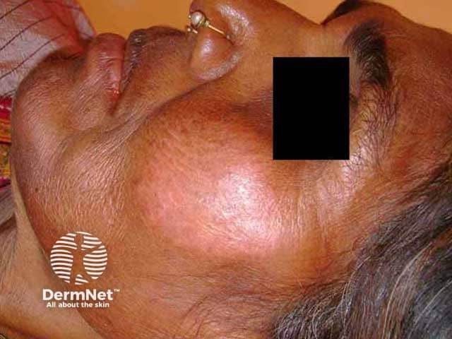
The clinical manifestations of leprosy depend on the immune response to M. leprae/lepromatosis and range over a spectrum from multibacillary lepromatous leprosy (LL) showing limited or low immunity to M. leprae/lepromatosis, to paucibacillary tuberculoid leprosy (TT) with a strong immune response.

What are the clinical features of leprosy?

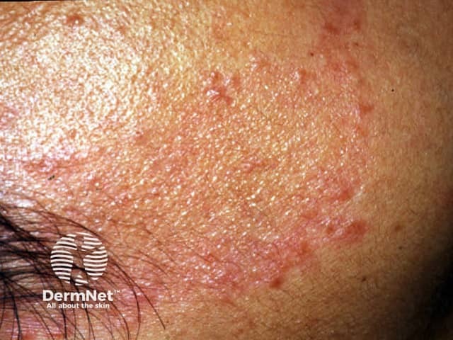
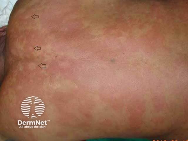
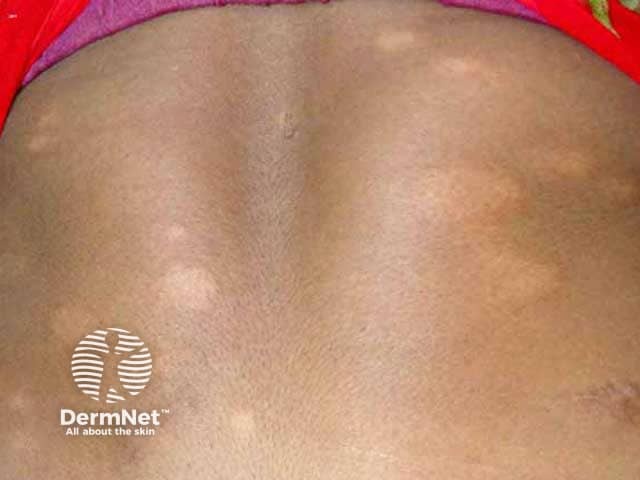
In over 90% of patients, the first symptom noticed is numbness. Temperature is the first sensation lost, followed by light touch, pain, and then deep pressure. This may precede the development of cutaneous lesions by years. The initial skin lesions are usually of the indeterminate type, presenting as a solitary or small number of hypopigmented patches before evolving into borderline tuberculoid or lepromatous types.

Tuberculoid leprosy

Tuberculoid (TT) leprosy is the paucibacillary form defined clinically by:

  • A few (1–2) sharply defined red patches with raised borders or a single larger hypopigmented patch less than 10 cm in diameter
  • Loss of sweating with rough dry hairless skin in the patches
  • Loss of sensation in lesions
  • Affected nerves are thickened and tender on palpation.

Tuberculoid leprosy

Borderline tuberculoid leprosy

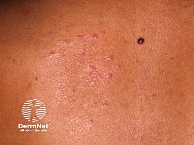
Borderline tuberculoid (BT) leprosy presents with:

  • Similar lesions to TT but larger in size, more numerous (5-20), and can be less well-defined
  • Asymmetrical distribution
  • Satellite lesions
  • Anaesthesia over the lesions is less pronounced compared to TT
  • Peripheral nerves are affected in an asymmetrical pattern and can cause deformity and disability.

Borderline tuberculoid leprosy

Borderline borderline leprosy

Borderline borderline (BB) leprosy is a rarely seen, transient, unstable form of leprosy defined by:

  • Multiple lesions of varying size, shape, and distribution
  • Skin-coloured or erythematous lesions
  • Characteristic, but rare, inverted saucer-shaped lesions with sloping edges and punched out centre (Swiss cheese lesion).

Borderline borderline leprosy

Borderline lepromatous leprosy

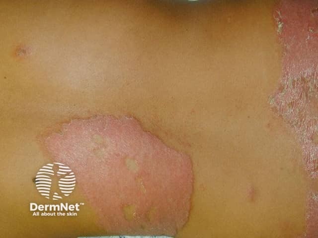
Borderline lepromatous (BL) leprosy is characterised by:

  • Widespread bilaterally symmetrical lesions
  • Macules, papules, and nodules of variable size and shape
  • Sensation and hair growth remain normal within a lesion
  • Characteristic glove and stocking numbness
  • Widespread peripheral nerve involvement.

Borderline lepromatous leprosy

Lepromatous leprosy

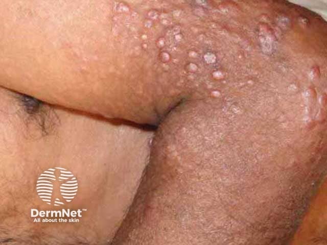
Lepromatous (LL) leprosy is the multibacillary form defined by:

  • Early symptoms of nasal stuffiness, discharge, and bleeding
  • Swelling and thickening of limbs, especially ankles and legs with subsequent ulceration
  • Widespread poorly defined hypopigmented and erythematous macules with a shiny surface and normal sensation
  • Progression to widespread infiltration of skin forming nodules and plaques
  • Characteristic leonine facies with thickening of the forehead, loss of eyebrows and eyelashes (madarosis), distortion of the nose, and thickening of the earlobes
  • Involvement of other systems:
    • Eyes — corneal anaesthesia, keratitis, corneal ulceration, uveitis, glaucoma, irreversible blindness
    • Testes — orchitis, testicular atrophy, sterility
    • Liver — hepatitis, hepatic amyloidosis
    • Kidneys — glomerulonephritis, renal amyloidosis
    • Bones — osteoporosis, resorption of digits.

Lepromatous leprosy

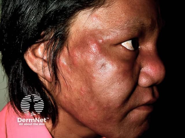
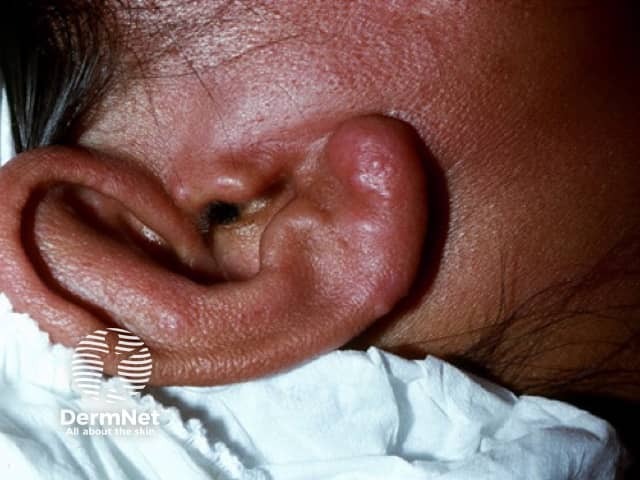
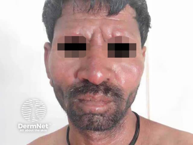
A number of clinical variants of lepromatous leprosy are described.

  • Histoid leprosy — multiple dome-shaped, skin-coloured to coppery-red papules and nodules (histoid lepromas) of variable size mainly on the limbs and trunk, or along peripheral nerves particularly in males. This has a characteristic histology, and typically erupts during treatment of LL reflecting treatment-resistant bacilli or focal loss of immunity. There is concern this form of leprosy may act as a reservoir making eradication difficult.
  • Lucio leprosy (diffuse lepromatous leprosy) — presents as a smooth infiltration of the skin, especially on the face and hands. It is particularly seen in Central and South America where it is called 'lepra bonita' (pretty leprosy).
  • Verrucous lepromatous lesions — are filiform, horn-like, or fissured hyperkeratotic warty projections or plaques on the distal lower limb and feet. It is a rarely described presentation seen in advanced lepromatous leprosy.

Histoid leprosy

Pure neural leprosy

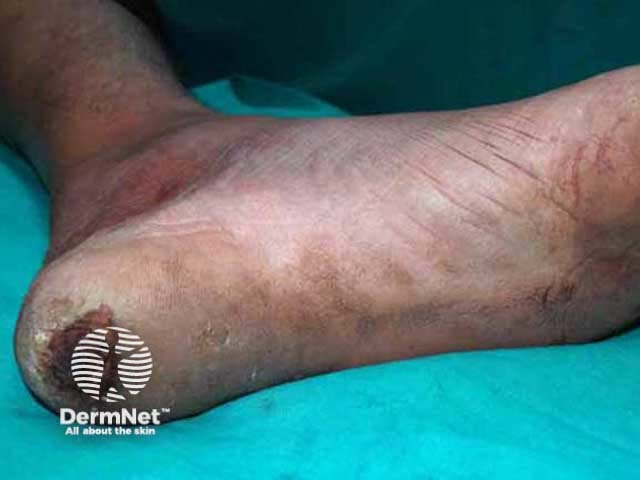
Pure neural leprosy (PNL) is common in India and Nepal. It presents with only peripheral nerve tenderness and thickening without skin lesions. However the nerve damage can result in loss of sensation and hence trophic ulcers. Diagnosis is difficult and requires nerve biopsy.

Pure neural leprosy

What are the complications of leprosy?

Lepra reactions occur in 30–50% of patients with leprosy. They may occur before, or more often, after the start of treatment. These are sudden responses resulting from the release of immunologically active bacilli or its products leading to localised or systemic symptoms and signs. Such reactions are responsible for most of the nerve damage, deformity, and disability.

How is leprosy diagnosed?

Leprosy has very characteristic clinical features, and dermoscopy is being used more often to aid clinical diagnosis.

Diagnosis should be confirmed by one of the following investigations.

  • Skin Slit Smear — a small slit is made using a sharp blade over the skin of the earlobe, forehead, or lesional skin, then a smear is made by scraping the exposed dermis onto a glass slide and examining for acid fast bacilli under microscopy; useful for multibacillary leprosy only.
  • Lepromin test — is an intradermal test for delayed type hypersensitivity to M. leprae antigens; although not specific, it is helpful for classifying the type of leprosy.
  • Skin biopsy — may show typical features, depending on the type of leprosy (see Leprosy pathology); special stains may be required to demonstrate the bacilli.
  • M. leprae DNA PCR is very specific for detecting leprosy organisms.

What is the differential diagnosis for leprosy?

The potential differential diagnosis list for leprosy is long and depends on the type and clinical features.

What is the treatment for leprosy?

The treatment of leprosy aims to stop active infection and minimise complications and deformity. Residual disabilities may require corrective reconstructive surgery to allow day-to-day activity.

Most endemic countries follow the WHO recommended multi-drug therapy (MDT) of antibiotics; the combination of drugs selected and duration of treatment depends on the type of leprosy. The 2018 WHO guidelines recommend three drugs including clofazimine for 6 months in paucibacillary leprosy and 12 months for multibacillary disease.

First-line antibiotics used in the treatment of leprosy are dapsone, rifampicin and clofazimine. Other drug options include ofloxacin, moxifloxacin, minocycline, clarithromycin, rifapentine, and diarylquinolone. Vaccines and other forms of immunotherapy are being trialled.

What is the outcome for leprosy?

Once appropriate treatment has been commenced, the skin lesions slowly subside. However, nerve damage cannot be reversed.

Can leprosy be prevented?

Post-exposure prophylaxis using rifampicin reduces the development of paucibacillary leprosy by 50%.


Bibliography

  • da Silva MB, Portela JM, Li W, et al. Evidence of zoonotic leprosy in Pará, Brazilian Amazon, and risks associated with human contact or consumption of armadillos. PLoS Negl Trop Dis. 2018;12(6):e0006532. doi:10.1371/journal.pntd.0006532. PubMed Central
  • Fischer M. Leprosy — an overview of clinical features, diagnosis, and treatment. J Dtsch Dermatol Ges. 2017;15(8):801–27. doi:10.1111/ddg.13301. PubMed
  • Jopling WH, McDougall AC. Handbook of Leprosy, 5th edn. New Delhi: CBS Publishers & Distributors, 1996.
  • Kar HK, Kumar B (eds). IAL Textbook of Leprosy. JAYPEE Brothers Medical Publication, 2010.
  • Maymone MBC, Laughter M, Venkatesh S, et al. Leprosy: clinical aspects and diagnostic techniques. J Am Acad Dermatol. 2020;83(1):1–14. doi:10.1016/j.jaad.2019.12.080. PubMed
  • Maymone MBC, Venkatesh S, Laughter M, et al. Leprosy: treatment and management of complications. J Am Acad Dermatol. 2020;83(1):17–30. doi:10.1016/j.jaad.2019.10.138. PubMed
  • Sharma R, Singh P, McCoy RC, et al. Isolation of Mycobacterium lepromatosis and development of molecular diagnostic assays to distinguish M. leprae and M. lepromatosis. Clin Infect Dis. 2019;ciz1121. doi:10.1093/cid/ciz1121. PubMed
  • White C, Franco-Paredes C. Leprosy in the 21st century. Clin Microbiol Rev. 2015;28(1):80–94. doi:10.1128/CMR.00079-13. PubMed
  • Yu R, Jarrett P, Holland D, Sherwood J, Pikholz C. Leprosy in New Zealand: an epidemiological update. N Z Med J. 2015;128(1414):9–14.

On DermNet

Other websites

Books about skin diseases


Related information

Sign up to the newsletter