DermNet provides Google Translate, a free machine translation service. Note that this may not provide an exact translation in all languages

Translate

Causes of atopic dermatitis

Author: Dr Amy Stanway, Department of Dermatology, Waikato Hospital, Hamilton, New Zealand, February 2004.


toc-icon

Introduction

There is no known single cause for atopic dermatitis (eczema) and it probably represents more than one condition. There are many theories regarding the underlying mechanisms. Current research is investigating the roles of the immune system, skin structural gene mutations, defects in the skin cells (keratinocytes), the skin surface microbiome (bacteria, viruses and yeasts), and many other factors.

Current theories identify that atopic dermatitis is primarily a disease of the immune system, with cytokines being critical components to the disease. These cytokines, particularly IL-4 and IL-13 (Th2 pathway cytokines) and IL-22 (the Th22 axis cytokine) cause barrier defects and inflammation that result in the clinical features of eczema.

Atopic dermatitis

What is atopy?

Atopy refers to the tendency to asthma, eczema and hay fever. Atopy is mostly inherited (genetic). It is characterised by an overactive immune response to environmental factors. The same factors have no effect on the skin of a non-atopic. Yet, despite their genetic background, some children from an atopic family never develop atopic dermatitis and children with no family history can suffer from it. For more information, see the webpage on atopy.

Atopic dermatitis is a disease of the whole body that manifests in the skin. Events that upset the body in other ways (such as viral infection, teething, eating certain foods) may have an effect on dermatitis. Flares may also occur without obvious provocation and can be very frustrating.

The inherited barrier defect

There is emerging evidence that inflammation in atopic dermatitis is associated with immune-mediated and inherited abnormalities in the skin barrier. This barrier failure causes increased permeability of the skin and reduces its antimicrobial function.

The main inherited abnormality causing disordered barrier function is filaggrin expression. Filaggrins are filament-associated proteins which bind to keratin fibres in the epidermal cells. The gene for filaggrin (FLG) resides on chromosome 1 (1q21.3). This gene was first identified as the gene involved in ichthyosis vulgaris. Abnormal filaggrin is associated with early-onset, severe and persistent atopic dermatitis.

It is postulated that the loss of filaggrin results in:

  • Corneocyte deformation (flattening of surface skin cells), which disrupts the organisation of the extracellular lipid (fat) — the lamellar bilayers.
  • A reduction in natural moisturising factors, which include metabolites of pro-filaggrin.
  • An increase in skin pH which encourages serine protease activity — these are enzymes which digest lipid-processing enzymes and the proteins that hold epidermal cells together. Serine proteases also generate active cytokines like IL-1a and Il-1beta and promote skin inflammation.

Proteins under investigation in atopic eczema include structural compounds, such as hornerin, cornulin, claudin 1/23 and ceramides, enzymes, such as kallikrein (a protease) and serine peptidases. 

The immune system

The immune system develops in the first six months of life. There is a generally an equilibrium of the two main types of T Helper lymphocytes (small white blood cells), Th-1 and Th-2. In atopic dermatitis, there is often an imbalance, with far more Th-2 cells and their associated chemical messengers (cytokines). In some children, there are also high levels of the antibody immunoglobulin E (IgE) antibodies and eosinophils (the white blood cells associated with allergy). 

The Th2 associated cytokines contribute to the loss of skin barrier function:  

  • Water is lost
  • Irritants may penetrate (soap, detergent, solvents, dirt etc.)
  • Allergens may penetrate it (pollens, dust-mite antigens, microbes).

The specialised immune cells of the epidermis (Langerhans cells) have an increased response to these antigens in atopic dermatitis and interact with dermal T cells to produce an even greater Th2 response further exacerbating the barrier defect.

  • Ceramide (a fatty acid) is reduced
  • Filaggrin is reduced
  • Antimicrobial peptides are reduced
  • Bacteria colonise and infect the skin
  • Infections are more difficult to control

So both the genetic make-up of the individual and external environmental factors contribute to the likelihood of developing eczema, its severity and its response to treatment.

Very rarely, atopic dermatitis may be due to an underlying inherited immune deficiency such as Job syndrome. In this disease, dermatitis appears very soon after birth and is complicated by severe infections.

The microbiome

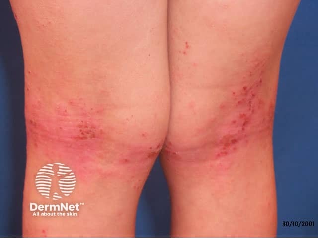
The functions of microbial components of the skin flora have only recently become of interest to atopic dermatitis researchers. It is postulated that different organisms in different body sites might account for the characteristic distribution of active lesions of atopic dermatitis — for example in the moist elbow and knee creases. 

The microbiome likely contributes to normal and abnormal inflammatory responses in the skin.  Flares of atopic dermatitis are accompanied by a proliferation of Staphylococcus aureus on lesional skin and a reduction in the biodiversity of the cutaneous microbiome (dysbiosis), with fewer malassezia yeasts and cutibacteria than are present on normal skin. Researchers report that emollients can increase biodiversity in eczema skin.

Another focus of research is the role of the intestinal microbiome.

What makes the skin get drier?

Dry skin is a sign of the loss of barrier function. Factors that make the skin even drier may make eczema harder to control:

  • Winter weather
  • Frequent washing particularly with very hot water
  • Washing in hard water (which increases pH)
  • Soap (which also increases pH) and antiseptics
  • Low humidity
  • High ambient temperatures
  • Chlorine in swimming pools

A brief shower or bath once a day is enough if you have dry skin. However, your doctor may specifically recommend bathing for longer or more often during the acute blistered phase of eczema. Use a non-soap cleanser.

Regular use of an emollient helps skin retain moisture and combat dryness.

What is the role of irritants?

Most people with eczema will notice that certain things seem to irritate their skin with immediate stinging or itching and may also cause a flare of eczema (irritant contact dermatitis). These are not allergies.

  • Soap
  • Harsh clothing detergents
  • Coarse fibres (wool and synthetic) and seams in undergarments
  • Cosmetic and perfumes
  • Prescribed and over-the-counter treatment creams
  • Dusty environments

It is common sense to avoid the substances that irritate.

  • Dilute washing powder: use a small amount in each load and make sure the clothes are well rinsed out in freshwater. Select non-fragranced laundry detergent.
  • If you use soap to wash your hands, rinse off thoroughly.
  • Wear gloves and covering clothing to protect against detergents, cleaning chemicals, solvents etc. at work and at home.
  • Limit preparations applied to the skin to those specifically prescribed for eczema.

The role of infection

Antigen stimulation

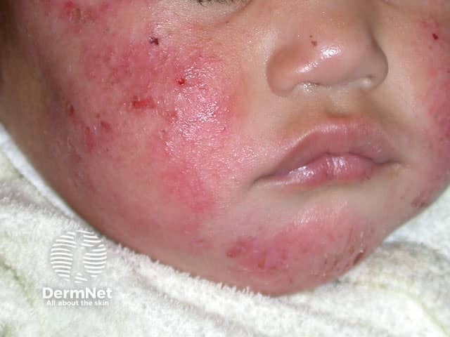
Infective organisms play an important role in triggering and aggravating atopic dermatitis. Bacteria (particularly staphylococci) and in some patients yeasts (malassezia and candida) contribute to chronic inflammation.

Bacterial infection

People who have atopic dermatitis are particularly prone to skin infections with Staphylococcus aureus.

  • This is in part due to breaks in dry, split skin, from scratching and from diminished barrier function.
  • They also seem to have a reduced ability to fight these common organisms.
  • The bacteria that cause infection are also commonly found on healthy skin. The oil on healthy skin is a protective mechanism against invasive infection because the bacteria are lipophobic (literally 'afraid of fat'). Staphylococcal bacteria thrive and invade dry, atopic skin.
  • Antimicrobial peptides on the skin surface normally fight these bacteria but may be deficient in the stratum corneum of atopic dermatitis.

As a result, people with atopic dermatitis frequently suffer from boils, folliculitis and infected eczema.

The infection causes eczema to worsen and become more resistant to the usual treatment with emollients and topical steroids. S. aureus produces enterotoxin. This induces the production of enterotoxin-specific IgE, resulting in proliferation and recruitment of more T cells and aggravating dermatitis.

Antibiotics are often required to eliminate the infection and control eczema. However, antibiotics may reduce the biodiversity of the microbiome.

Viral infections

Fungal infections

  • Some adults have a flaky pattern of eczema on the face and neck, which is related to skin colonisation by malassezia. In these patients, prick tests to malassezia may be positive and malassezia-specific IgE may be detected on blood testing. Eczema can improve with topical antifungal or oral antifungal treatment (itraconazole or ketoconazole). Intermittent antifungal treatment (e.g., once weekly) is often required long term.
  • Candida (thrush) is also more likely to thrive in poorly controlled, moist areas of eczema.

Dermatophyte infections (tinea) do not appear to be more prevalent in atopic dermatitis.

Allergens and eczema

  • There are many misconceptions and controversies regarding eczema and allergy. People who have atopic dermatitis associated with elevated IgE are likely to have allergies to food or environmental factors such as grass, cat dander and dust mites. However these allergies are often independent of their eczema, that is, even though the patient has a positive allergy test, exposure to the allergen does not directly affect the severity of eczema. 
  • Current research indicates that skin barrier defects that occur in atopic dermatitis alter immune function and so can induce food allergy secondarily.  
  • It is common for prick tests for allergy to be positive in atopic individuals because of the extreme sensitivity of eczematous skin to any insult. The tests are done by scratching the skin with a small amount of a potential allergen such as cat dander. In people with eczema scratching the skin may cause a raised mark, i.e. a positive test result even without any allergen (this is called a false positive). This means the test is unreliable for diagnosing allergies in those with eczema.
  • Blood tests (RAST) measure the level of specific IgE to different allergens. They also have a high false-positive rate and may not reflect the allergen's effect on atopic dermatitis.
  • If a true allergy is present AND exposure to the allergen results in a persistent worsening of eczema, removal of the allergen from the affected patient's environment can result in improvement of eczema.

Food allergies

Food allergies affect about a third of children with eczema. They are most commonly to egg, cows milk, soy, wheat, peanuts and fish. The reaction may be acute urticaria (hives) sometimes accompanied by swelling of the face and tongue (angioedema) or abdominal pain shortly after ingesting the offending food. Severe allergy causes anaphylaxis and the patient may collapse and even die. Any tiny amount of food allergen can cause this response. These reactions are not eczema.

However, eczema can be aggravated by certain foods in other ways (food intolerance).

  • Some foods, particularly fruits, contain salicylates. Salicylates can increase histamine release and thus cause temporary itching and redness. This is not an allergy.
  • Parents often suspect food allergies in children with atopic dermatitis because they notice eczema getting worse when new foods are introduced. Whilst it is very common for eczema to become more difficult to manage when new foods are being introduced it is only very rarely due to a true allergy. As eczema tends to reflect other stresses the body is facing, any new challenge, such as the digestive system getting used to a new food, may cause a temporary increase in eczema symptoms. Just as the gut takes some time to tolerate certain new foods, so does the skin.
  • The increase in eczema symptoms tends to improve after a month or so of the new food. Infants and children are constantly trying new foods, therefore, this difficulty with eczema can last for several months. As a wide variety of foods are very important for young children, it is worth persevering with the increased demands of eczema management during this period. Restricting foods excessively and unnecessarily may cause more detriment to the child's health than a transient worsening of eczema.
  • If you have a strong suspicion of a food allergy, consult an allergist for advice. Consult a registered dietician to make sure any restrictive diet remains nutritionally sound.

Environmental allergies

Environmental allergens may sometimes be related to atopic dermatitis. Many children with atopic dermatitis are allergic to grass, dust mites and cat dander. Usually, this manifests as an immediate reaction with a runny nose, sneezing and swollen eyes, and improves after removal from the allergy source. It is uncommon for this type of allergy to cause persistent worsening of eczema.

  • A positive test result to an environmental allergen may prove unhelpful in the management of eczema. This is in part due to the high rate of false-positive reactions as explained above and in part because avoidance of these allergens is very difficult, if not impossible.
  • Exposure to dust mites can be reduced, but not eliminated, by regularly vacuuming the house, keeping soft toys and clothes shut away in cupboards, limiting floor carpets, washing linen in hot water and using a protective mattress and pillow covers.
  • Grass pollen is very difficult to avoid unless the child is forbidden to play outside.
  • The relationship between cat dander allergy and eczema is controversial. There is some evidence to suggest that having more cats in the house early in life protects children from developing atopy (asthma, eczema and hay fever). It is not clear at this time whether those with cat dander allergy should be advised to get rid of their cat or not.

Allergic contact dermatitis is equally common in those without atopic dermatitis.

Stress

  • Adults and children are liable to experience an exacerbation in eczema secondary to physical, mental or social stress.
  • Stress can come in many forms. Any other illness including a mild common cold may cause a flare in eczema.
  • Social stress such as a move to a new area, change in school, family conflict etc. may also affect eczema.
  • Eczema may be the source of stress: it is uncomfortable and can be a major cosmetic concern. The cost of medications and time off work for the patient and caregiver can also be very stressful.

Climate

  • Climate undoubtedly plays a major role in the severity of eczema. Cold, damp climates may cause eczema to become more resistant to treatment. Keeping the temperature even throughout the house is probably helpful. Dehumidifiers tend to be more beneficial for asthma than for eczema as low environmental humidity can dry out the skin further.
  • Moving to a new house or new area can have a beneficial or detrimental effect on eczema.
  • Most people find that their eczema is better in the summer months. This is partly due to the increased exposure to ultraviolet light, which (in moderation) can have a beneficial effect on eczema. Sunburn is never a good idea! A small percentage of eczema sufferers find that the sun makes their eczema worse and should protect from sun exposure as much as possible. This is called photosensitive eczema.
  • Most people with eczema will become itchier and have redder skin when they are hot. Keeping cool in hot environments using a fan or air conditioning, and having cool showers or baths will help.
  • Choose superfine natural fibre clothing and bed linen to reduce sweating and help keep skin cool. Avoid coarse wool underlays.


References

  • Elias PM and Steinhoff M, “Outside-to-Inside” (and Now Back to “Outside”) Pathogenic Mechanisms in Atopic Dermatitis, Journal of Investigative Dermatology (2008), 128, 1067–1070.
  • Elias PM et. al Basis for the barrier abnormality in atopic dermatitis: Outside-inside-outside pathogenic mechanisms. J Allergy Clin Immunol 2008;121:1337–43.
  • Bieber T, Atopic Dermatitis, N Engl J Med 2008;358:1483–94.
  • Avgerinou G et. al. Atopic dermatitis: new immunologic aspects, International Journal of Dermatology 2008, 47, 219–24
  • Morar N et. al. Filaggrin Mutations in Children with Severe Atopic Dermatitis, Journal of Investigative Dermatology (2007) 127, 1667–72;
  • Vickery B, Skin barrier function in atopic dermatitis, Curr Opin Pediatr 19:89–93. 2000
  • Sandilands A, et al. Filaggrin’s Fuller Figure: A Glimpse into the Genetic Architecture of Atopic Dermatitis. Journal of Investigative Dermatology (2007) 127, 1282–4.
  • Dybboe R, Bandier J, Skov L, Engstrand L, Johansen JD. The Role of the Skin Microbiome in Atopic Dermatitis: A Systematic Review. Br J Dermatol. 2017 Feb 16. doi: 10.1111/bjd.15390. [Epub ahead of print] Review. PubMed PMID: 28207943. PubMed
  • Williams MR, Gallo RL. The role of the skin microbiome in atopic dermatitis. Curr Allergy Asthma Rep. 2015 Nov;15(11):65. doi: 10.1007/s11882-015-0567-4. Review. PubMed PMID: 26404536. PubMed

On DermNet

Other websites

Books about skin diseases


Related information

Sign up to the newsletter