Introduction
Demographics
Causes
Clinical features
Variation in skin types
Complications
Diagnosis
Differential diagnoses
Treatment
Prevention
Outcome
Chronic actinic dermatitis (CAD) is a rare, immune-mediated photodermatosis characterised by abnormal photosensitivity to ultraviolet (UV) light and occasionally to visible light. It manifests as a dermatitis affecting predominantly, but not exclusively, photo-exposed sites.
The condition is also known as chronic photosensitivity dermatitis and actinic reticuloid.


In a retrospective study of 130 CAD patients, 36.2% were diagnosed under the age of 40 years. An earlier onset has been linked with:
The pathogenesis of chronic actinic dermatitis is not fully understood, but it is thought to be immune-mediated.
CD8+ T cells have been identified in the dermis of lesional skin in CAD, similar to what is observed in allergic contact dermatitis. It has been hypothesised that CAD is a delayed (type IV) hypersensitivity reaction to an unknown, endogenous photo-induced antigen — a molecule that has become immunogenic by exposure to light.
An alternative hypothesis is that CAD results from cross-reactivity between an endogenous (self) antigen and a photoallergen. The photoallergen triggers an allergic response, and the self-antigen allows this response to persist even in the absence of the offending photoallergen.
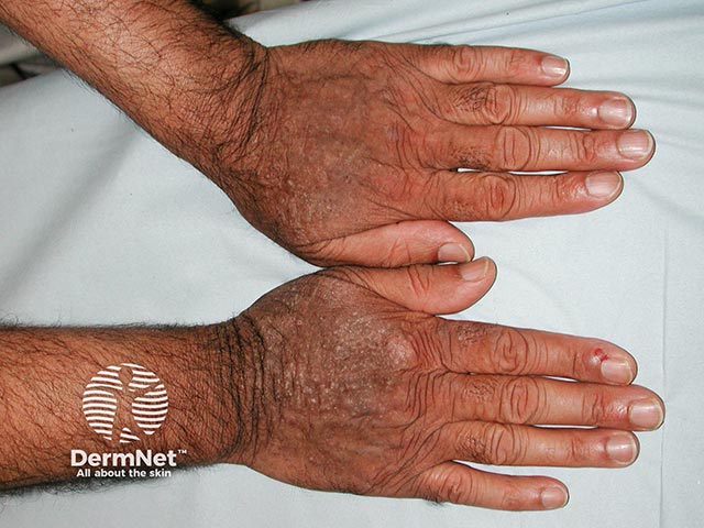
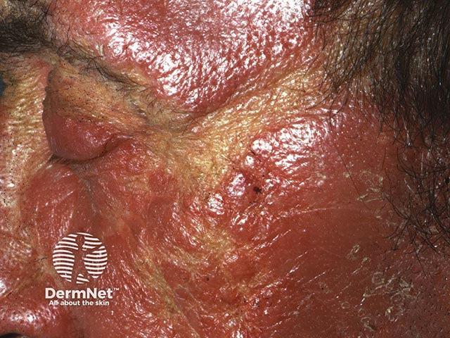
Chronic actinic dermatitis presents with an intensely pruritic, eczematous rash that classically involves photo-exposed sites. The face, upper chest, neck and dorsal aspects of the hands and arms are commonly affected.
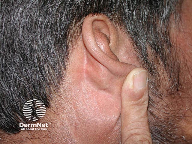
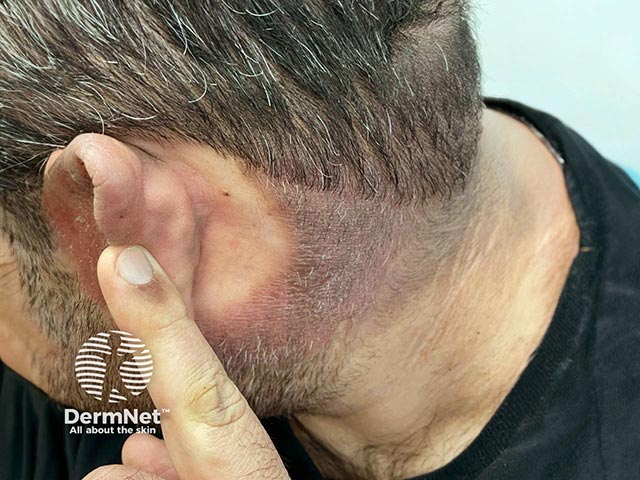
There is often notable sparing of the sun-protected areas such as the upper eyelids, postauricular areas, submental chin, and nasolabial folds. In severe cases and in patients with coexistent atopic dermatitis or allergic contact dermatitis, CAD can generalise to affect even sun-protected sites. Very rarely patients may present with erythroderma.
For some individuals, even brief sun exposure—as little as under a minute—may be sufficient to trigger dermatitis within 72 hours. In the most severe cases, this may remain active year-round with loss of the typical seasonal variation. Patients are at risk even on dull days and through window glass. Some people also react to artificial light sources, especially naked fluorescent lamps.
Actinic reticuloid is a severe variant of chronic actinic dermatitis in which patients present with thickened nodules and lichenified plaques in a similar photo-distribution.
In general, darker skin types present with less pronounced erythema. Other clinical features of chronic actinic dermatitis and associated range of UV-sensitivity do not vary according to skin type. However, the average age of onset may be earlier in darker skin types.
Initial concerns of increased risk of lymphoma transformation have been disproven.
Phototesting should be performed in all suspected cases of chronic actinic dermatitis to confirm the diagnosis. This involves exposing the skin to controlled amounts of light at specific wavelengths. The reactions that develop confirm the presence of an abnormal reaction to the light/radiation.
A monochromator will elicit an erythematous or eczematous response to low levels of ultraviolet (UV) radiation (and sometimes visible light). Photosensitivity to either UVB alone, UVA and UVB, or, less commonly, UVA alone are typically seen. However, the existence of CAD with pure UVA sensitivity is disputed by some photodermatologists and requires careful exclusion of other causes, particularly drug-induced photosensitivity. In severe cases, sensitivity may extend to include visible light.
A solar simulator or broad-spectrum UVA/UVB source may also be used to provoke a reaction for diagnostic purposes.
Patch and photopatch testing are also used to assess for relevant contact and/or photo-contact allergens, as guided by the clinical history.
Lupus serology (antinuclear antibodies and extractable nuclear antigens) is typically requested to rule out other conditions that may rarely mimic CAD.
Investigations to exclude cutaneous T-cell lymphoma may be performed, particularly in actinic reticuloid variants where severe lichenification can produce pseudolymphomatous plaques.
Vitamin D levels should be assessed and managed accordingly, given that photoprotection is an essential part of CAD management.
Strict photoprotection is the cornerstone of management for chronic actinic dermatitis. In severe cases, it may be necessary to admit the patient to a dark room in the hospital.
Patients should be advised to:
Strict photoprotection is the most important preventative measure. Avoidance of known contact or photocontact allergens also has a role in preventing symptom recurrence.
For most people, chronic actinic dermatitis is a lifelong condition that requires significant lifestyle changes to avoid sunlight as well as contact allergens.
However, CAD can spontaneously resolve, sometimes many years after its onset. A cohort study reported a 20% probability of resolution of abnormal photosensitivity within 10 years after diagnosis. In the same cohort, identification of separate contact allergens in two or more patch tests predicted a poorer prognosis for resolution.

