Introduction
Demographics
Causes
Clinical features
Complications
Diagnosis
Differential diagnoses
Treatment
Outcome
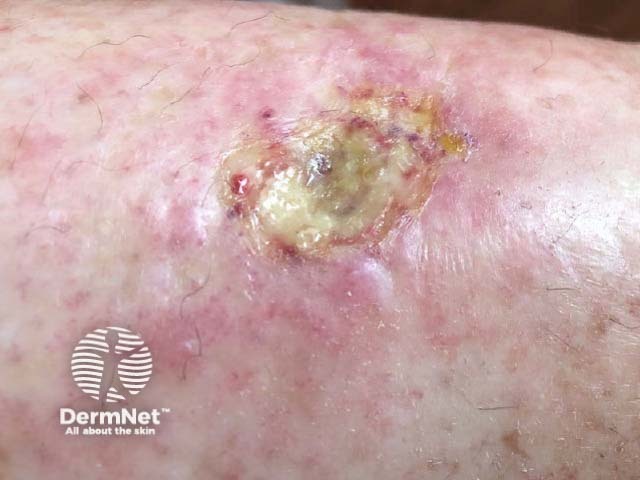
Hydroxyurea-induced cutaneous ulcer is a painful ulcer on the lower leg of a patient on high-dose long-term treatment with hydroxyurea. Ulcers are a common adverse cutaneous effect, reported in up to 10% of patients undergoing long-term hydroxyurea treatment for chronic myeloproliferative disorders.
Hydroxyurea-induced cutaneous ulcers develop in middle-aged or elderly patients (median age 67 years) with chronic myeloproliferative disorders actively being treated and maintained on hydroxyurea. The ulcers typically develop after 5 years or more of treatment. A female predominance (61%) has been reported. The most commonly associated myeloproliferative disease reported has been essential thrombocythaemia. Arterial and venous insufficiency may be present, but is not a requirement.
Hydroxyurea is a cytostatic agent that inhibits ribonucleotide reductase in the S (synthesis) phase of the cell cycle.
A cumulative toxic effect is believed to be involved in the development of the ulcer. Ulcer formation during hydroxyurea treatment is likely to be multifactorial in origin, including due to cytotoxic effects on epidermal basal keratinocytes and vascular endothelial cells, inhibition of collagen formation, and disturbance of the microvascular circulation.
It is not known whether hydroxyurea alone is responsible for the ulcer, or whether the underlying myeloproliferative disorder also contributes to the ulcer formation via effects on arterial and venous circulation leading to tissue ischaemia and delayed wound healing.
Trauma may play an initiating role given the common localisation around the malleoli and pretibial sites.
A patient with a hydroxyurea-induced cutaneous ulcer usually has a history of being on hydroxyurea for a few years before the ulcer develops. The median time from starting hydroxyurea treatment to ulcer diagnosis is 60 months. The typical hydroxyurea dose is 1g daily.
Other dermatological adverse effects of hydroxyurea may be noted.
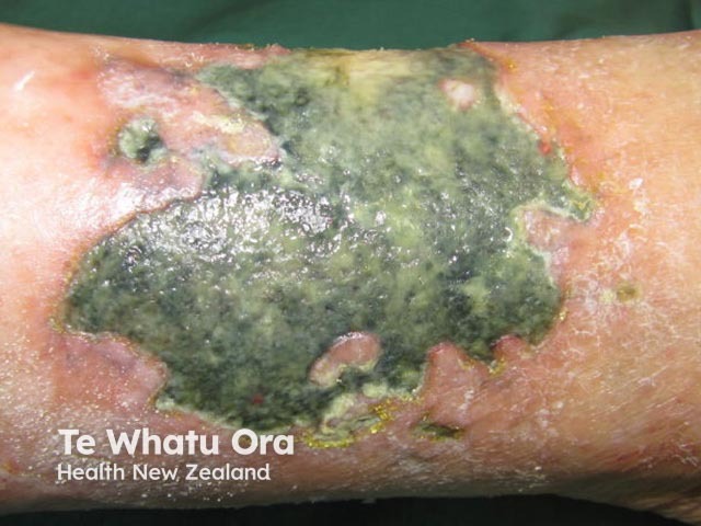
Secondary bacterial infection of the ulcer is the most common complication. This should be suspected if cellulitis occurs around the ulcer. However, chronic leg ulcers are often colonised by bacteria, and antibiotics are not indicated for localised slough or purulence, which is a sign of bacterial biofilm.
Healing can be delayed or incomplete for hydroxyurea-induced skin ulcers even after cessation of the hydroxyurea.
Analgesia required for the ulcer pain can have adverse effects in this predominantly elderly population.
The diagnosis of hydroxyurea-induced cutaneous ulcer is clinical, based on history and examination. The patient should be assessed for vascular disease (venous, arterial, or small vessel disease), which can be co-existent.
Histopathological features of hydroxyurea-induced cutaneous ulcer are non-specific and include epidermal atrophy and dermal sclerosis without inflammation.
Diagnosis is often missed or delayed due to the non-specific features.
The differential diagnosis of hydroxyurea-induced ulcer includes all forms of chronic leg ulcer. Most leg ulcers involve a component of vascular disease contributing to the development or poor healing of the ulcer.
A venous stasis ulcer is not usually painful. There are associated features of venous stasis including swelling, haemosiderin staining, hyperkeratosis, and lipodermatosclerosis.
An arterial ulcer tends to be painful at night with elevation of the leg. Distal pulses may be reduced or absent.
A diabetic foot ulcer tends to be located over a pressure area such as the heel or tip of a toe in a diabetic patient with peripheral neuropathy and microvascular disease.
Cessation of hydroxyurea therapy is essential for ulcer healing to occur. Together with wound care, this leads to ulcer resolution in a majority of patients. It is typical of hydroxyurea-induced skin ulcers that healing is not seen with wound care alone.
There have also been case reports of successful treatment of recalcitrant hydroxyurea-induced foot ulcers with timolol.
Resolution of the ulcer usually occurs within three months of ceasing hydroxyurea, although complete healing may take up to 24 months.
A small proportion of patients experience only partial ulcer healing and improvement in symptoms despite ceasing hydroxyurea.
Patients restarted on hydroxyurea therapy after complete ulcer resolution may quickly redevelop ulcers. These recurring ulcers do not heal until hydroxyurea therapy is ceased again.

