DermNet provides Google Translate, a free machine translation service. Note that this may not provide an exact translation in all languages

Translate

Necrobiosis lipoidica

Author: Hon A/Prof Amanda Oakley, Dermatologist, Hamilton, New Zealand, 2006. DermNet NZ Update August 2021


toc-icon

What is necrobiosis lipoidica?

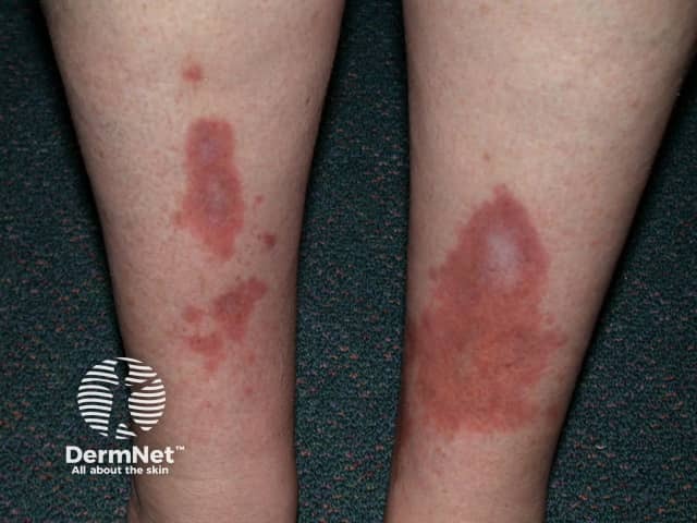
Necrobiosis lipoidica is a rare granulomatous skin disorder typically described on the shin of diabetics.

Necrobiosis lipoidica

Who gets necrobiosis lipoidica?

Necrobiosis lipoidica is three times more common in females than in males, and usually develops in young and middle-aged adults.

  • 1% of patients with diabetes will develop necrobiosis lipoidica.
  • It can occur in both type 1 and type 2 diabetes mellitus.
  • 11–65% of patients with necrobiosis lipoidica have diabetes or prediabetes.
  • Diabetes associated with necrobiosis lipoidica can be well controlled or poorly controlled.
  • Patients with type 1 diabetes are more likely to be younger than those with type 2 diabetes or without diabetes.
  • Other associations include obesity, hypertension, dyslipidaemia and thyroid disease.
  • Necrobiosis lipoidica is rare in children.

What is the cause of necrobiosis lipoidica?

The cause of necrobiosis lipoidica is unknown. Theories have included blood vessel changes such as develop in diabetes, or antibody-mediated vasculitis.

What are the clinical features of necrobiosis lipoidica?

Necrobiosis lipoidica begins as a dull red papule or plaque on the shin which slowly enlarges into one or more yellowish-brown patches with a red rim.

The patches:

  • Most often occur on both shins and are found in other sites only rarely
  • May be asymptomatic or tender
  • May be round, oval, or an irregular shape
  • Central atrophy - shiny, pale, and thinned, with prominent blood vessels (telangiectasia)
  • May demonstrate reduced sweating and sensation.

Dermoscopy of necrobiosis lipoidica

  • Yellow structureless background
  • Linear vessels with uniform branching
  • White linear streaks

How does necrobiosis lipoidica present in differing skin types?

In skin of colour, the patches and plaques of necrobiosis lipoidica may be hyperpigmented around the periphery with hypopigmentation developing centrally with the atrophy and telangiectases.

What are the complications of necrobiosis lipoidica?

  • Ulceration complicates 1/3 of cases of necrobiosis lipoidica, usually following minor injury to an established patch. The ulcer may be very painful or painless.
  • Ulcers due to necrobiosis lipoidica are at risk of secondary bacterial infection and delayed healing.
  • Squamous cell carcinoma has been reported to develop in ulcerated necrobiosis lipoidica.

Ulcerated necrobiosis lipoidica

What is the differential diagnosis of necrobiosis lipoidica?

How is necrobiosis lipoidica diagnosed?

Necrobiosis lipoidica is diagnosed clinically when typical.

A skin biopsy may be performed to confirm the diagnosis. The histopathology is characteristic; it shows a granulomatous inflammatory reaction around destroyed collagen (necrobiosis). [see Necrobiosis lipoidica pathology].

What is the treatment for necrobiosis lipoidica?

Not all cases of necrobiosis lipoidica require treatment and treatment is generally disappointing. The following treatments are sometimes effective:

What is the outlook for necrobiosis lipoidica?

Necrobiosis lipoidica is a chronic condition that may remain stable or slowly progress over years. Spontaneous resolution has been reported.


Bibliography

  • Peckruhn M, Tittelbach J, Elsner P. Update: treatment of necrobiosis lipoidica. J Dtsch Dermatol Ges. 2017;15(2):151–7. doi:10.1111/ddg.13186. Journal
  • Shrestha S, Spierings N, Marahatta S. Necrobiosis lipoidica: a case report with dermoscopic review. Clin Case Rep. 2021;9(3):1171–4.  doi:10.1002/ccr3.3713. Journal 
  • Uva L, Freitas J, Soares de Almeida L, et al. Squamous cell carcinoma arising in ulcerated necrobiosis lipoidica diabeticorum. Int Wound J. 2015;12(6):741–3. doi:10.1111/iwj.12206. Journal
  • Yahya H. Necrobiosis lipoidica in a Nigerian woman — report of a case. Niger J Clin Pract. 2019;22(11):1626–8. doi:10.4103/njcp.njcp_40_19. Journal

On DermNet

Other websites

Books about skin diseases


Related information

Sign up to the newsletter