Introduction
Demographics
Causes
Clinical features
Diagnosis
Treatment
Outcome
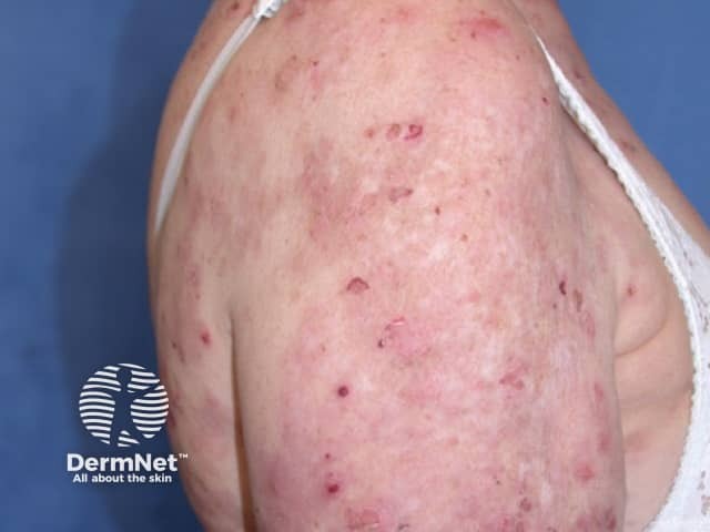
The pemphigus family are rare autoimmune blistering diseases affecting the skin and mucous membranes. Pemphigus foliaceus is a rare relatively benign form of pemphigus. In New Zealand pemphigus foliaceus is more often encountered than its more serious relative pemphigus vulgaris, although worldwide pemphigus vulgaris is more common. Pemphigus foliaceus is characterised by blistering lesions on otherwise healthy-looking skin. Blisters tend to form when the skin is rubbed (Nikolsky sign. There are currently six subtypes:
Pemphigus foliaceus affects people of all races, age and sex. It appears most commonly between the ages of 50-60 years.
Pemphigus foliaceus is an autoimmune disorder, which basically means that an individual's immune systems starts reacting against his or her tissue.
The building block cells of the epidermis are called keratinocytes. These cells are cemented together at special sticky spots called desmosomes. In pemphigus foliaceus, autoantibodies bind to a protein called desmoglein-1, which is found in desmosomes in the keratinocytes near the top of the epidermis. The result is the surface keratinocytes separate from each other, and are replaced by fluid: the blister. Because the blister is very close to the surface of the skin, the blisters rupture easily. In most cases, the autoantibodies are immunoglobulin type G (IgG) but in IgA pemphigus, the autoantibodies are type A (IgA).
Pemphigus foliaceus is sometimes provoked by sun exposure.
Endemic pemphigus foliaceus occurs in South America, where it is commonly known as Fogo selvagem. It appears to be set off by a virus transmitted by an insect bite.
Penicillamine, nifedipine, captopril, enalapril or nonsteroidal anti-inflammatory drugs most often provoke drug-induced pemphigus foliaceus. If the drug is stopped, there is a 50% chance the pemphigus foliaceus will clear up.
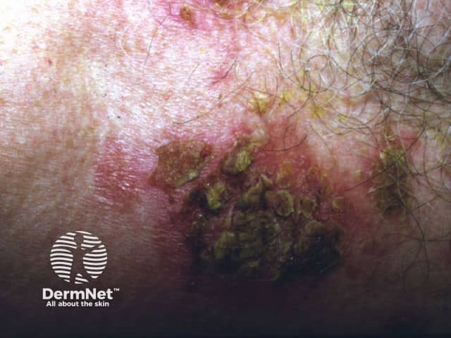
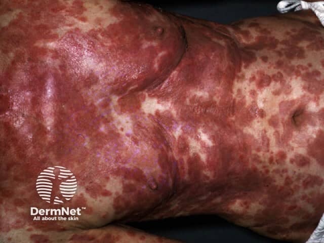
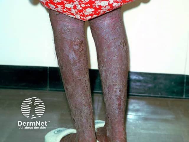
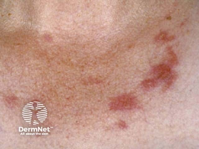
Pemphigus foliaceus produces superficial blisters confined to the skin, without involvement of mucous membranes. This is in contrast to pemphigus vulgaris where there may be extensive mucous membrane involvement (mouth, eyelids etc.).
The patient with pemphigus foliaceus is usually otherwise in good health. Small fluid-filled blisters first form on the trunk. Being superficial within the upper epidermis, they rupture very easily, and only erosions may be seen. On the face, scalp and upper trunk, the lesions are often scaly and crusty on a red and inflamed base. A burning sensation or localised pain may be felt.
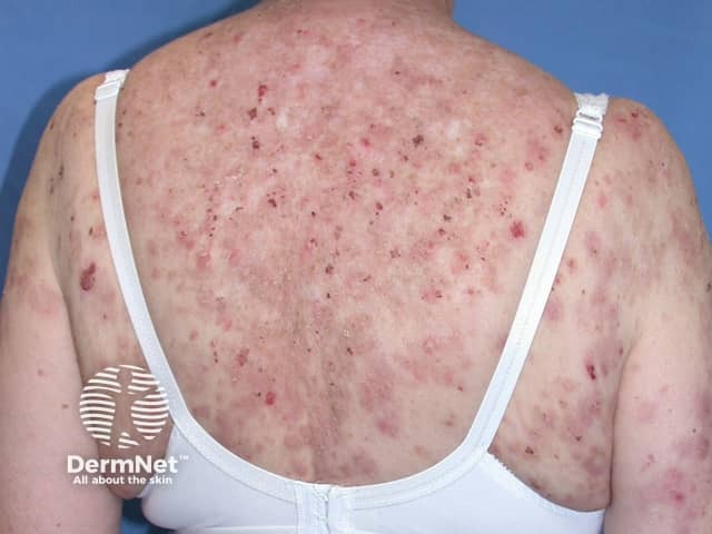
Pemphigus erythematosus (or Senear-Usher syndrome) is a variant of pemphigus foliaceus which shares clinical, histopathological, and serological features with systemic lupus erythematosus (SLE); they rarely coexist.
It accounts for 10% cases of all pemphigus disease. Clinically and immunologically, Senear-Usher syndrome shares features of SLE such as the presence of erythematous, scaly, and crusted plaques over the malar region and nose. Additionally, the disease may be worsened by exposure to sunlight as seen in SLE. Immunofluorescence demonstrates the presence of IgG and C3 at the dermo-epidermal junction (a feature distinguishing it from pemphigus foliaceus) in addition to intraepidermal IgG. Presence of anti-desmoglein (anti-dsg) 1 antibodies are seen and in 30-80% of patients; elevated ANA is also present.
The principles of management are the same as other forms of pemphigus, however it can be managed with lower steroid doses.
Diagnosis generally requires a skin biopsy, which shows typical features of rounded-up separated keratinocytes (called acantholytic cells) within the blisters in the upper layers of the epidermis.
Pemphigus foliaceus is confirmed by direct immunofluorescence staining of the skin sections to reveal antibodies.
In some cases, circulating antibodies can be detected by a blood test (indirect immunofluorescence test).
The primary aim of treatment is to prevent new areas from developing bacterial infections and promote healing of affected areas.
Topical treatment with corticosteroids is usually all that is necessary for mild pemphigus foliaceus. For more severe cases treatment is similar to that for pemphigus vulgaris. Oral antibiotics may be prescribed to treat secondary bacterial infection.
Remission off treatment may occur in some patients while in others, the problem may persist for several years.

