

Varicose veins — extra information
Introduction
Demographics
Causes
Clinical features
Complications
Diagnosis
Differential diagnoses
Treatment
Outcome
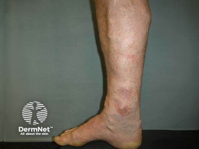
What are varicose veins?
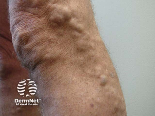
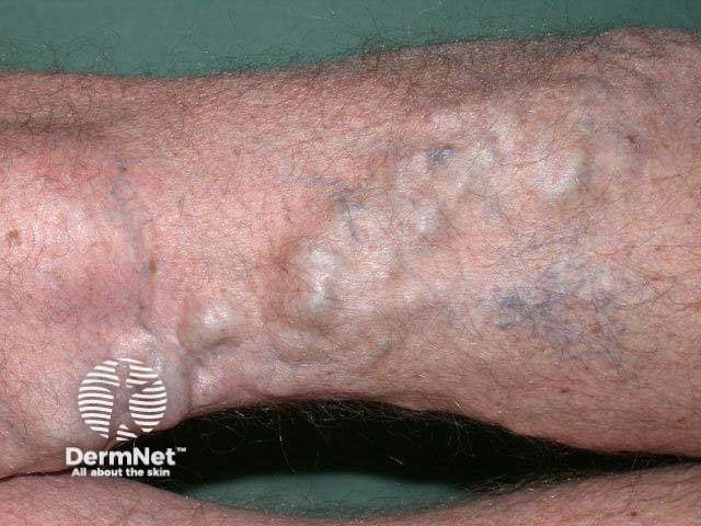
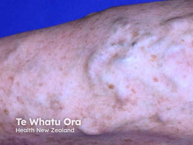
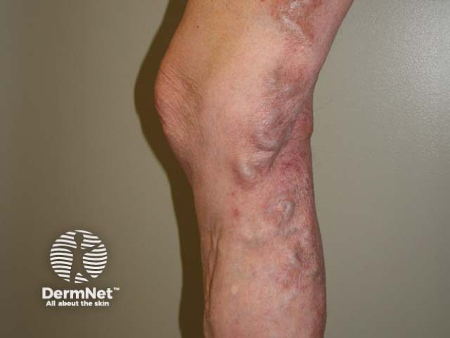
Varicose veins are engorged, tortuous, green, blue, or purple veins that are often found on the lower legs and feet.
Varicose veins are also called varices or varicosities.
Who gets varicose veins?
Approximately one-third of men and women aged 18–64 years have varicose veins [1]. They are more common in women and those with a family history of venous disease.
What causes varicose veins?
In normal leg veins, one-way valves direct the flow of venous blood from superficial venules to larger superficial veins, then to the deep veins, and eventually to the heart. Muscle contractions create a pumping action to help the flow of blood to the heart (known as the venous return). In the legs, varicose veins are a manifestation of venous insufficiency.
Risk factors for varicose veins
Risk factors for varicose veins include:
- Obesity — obesity increases venous reflux and venous pressure as a result of raised intra-abdominal pressure [2]
- Age — varicose veins become increasingly common with age [3]
- Pregnancy — the enlarged uterus causes increased intra-abdominal pressure and direct pressure on the iliac veins; hormonal changes also cause the valves and vessels to become more malleable during pregnancy [4]
- Prolonged standing — sustained pressure over time may cause venous distension and valve failure [3]
- A family history — primary valvular failure is hereditary, and varicose veins may affect both identical twins in 75% of cases [5].
What are the clinical features of varicose veins?
Patients with varicose veins present because they are unsightly and because of a feeling of discomfort, heaviness, itching, or a dull ache. Patients can also present with complications such as bleeding, ulceration, and thrombophlebitis.
What are the complications of varicose veins?
Bleeding
Superficial veins are prone to trauma and can bleed, which can be potentially life-threatening [6,7].
Ulceration
The increased pressure in varicose veins allows proteins, hormones, and circulating proinflammatory molecules to leak out of the vein and into the extravascular space, which leads to localised inflammation and the formation of a chronic venous leg ulcer.
Thrombophlebitis
Phlebitis is inflammation of a vein with erythema and painful hardening. It is often associated with a blood clot (thrombosis) that arises as a result of the sluggish circulation and factors that increase coagulation. Phlebitis may be superficial (ie, superficial thrombophlebitis) or deep (ie, deep venous thrombosis) [6].
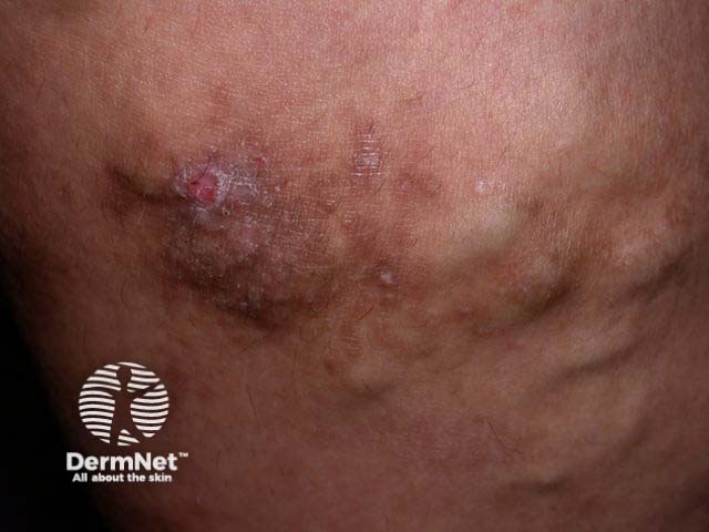
Venous stasis dermatitis
Venous stasis dermatitis is associated with increased venous pressure and pro-inflammatory molecules. Patients present with brown discolouration, pruritus, and discoid or circumferential, acute or chronic eczema on the distal lower limbs [7].
Lipodermatosclerosis
Lipodermatosclerosis is an inflammatory and thickened form of panniculitis resulting from the presence of pro-inflammatory molecules, poor venous outflow, and hypertension. The lower leg becomes reddened in acute lipodermatosclerosis and indurated in chronic lipodermatosclerosis.
How are varicose veins diagnosed?
Varicose veins are diagnosed clinically. A physical examination should include the entire venous system and it is usually conducted with the patient both lying down and standing up.
The UK's National Institute for Health and Care Excellence (NICE) uses the Clinical–Etiological–Anatomical–Pathophysiological (CEAP) classification of varicose veins.
CEAP classification
The CEAP classification categories are as follows:
- C0 — No visible or palpable signs of venous disease
- C1 — Telangiectasias or reticular veins
- C2 — Varicose veins; diameter > 3 mm
- C3 — Oedema
- C4 — Changes in skin and subcutaneous tissue, including pigmentation, eczema, lipodermatosclerosis, or atrophie blanche
- C5 — Healed venous ulcer
- C6 — Active venous ulcer [8].
A Duplex Doppler ultrasound assessment should be performed to determine the extent of disease and the level of truncal reflux (the failure of one of three of the main trunk veins) and to plan treatment options [8].
What is the differential diagnosis for varicose veins?
Varicose veins are larger than telangiectasia (small red 'thread' veins, < 1 mm in diameter) and venulectasia (blue reticular vessels, 1–3 mm in diameter). These are not detected on duplex ultrasound.
What is the treatment for varicose veins?
Weight loss (if overweight) and moderate physical activity should be encouraged in patients with varicose veins to reduce the risk of complications. Compression hosiery should be used to relieve discomfort and swelling, especially when travelling.
Treatment for varicose veins is available from a vascular service. Options for treatment are listed below.
Endovenous thermal ablation
Endovenous thermal ablation, using either a laser or radiofrequency device, causes an irreversible thermal injury in the vein wall, which leads to scarring and the absorption of the tissue over several months. The success rate for both methods of ablation is 95% [9,10].
Injection sclerotherapy
Sclerotherapy involves the injection of a sclerosant solution into the veins under ultrasound guidance to cause inflammation in the vessels and the eventual collapse of the varicose network. Smaller surface veins can be injected by microsclerotherapy, involving smaller gauge needles with smaller amounts of sclerosant [11].
Endovenous adhesive ablation
Endovenous adhesive ablation, or 'vein glue' technique, involves the injection of a medical-grade adhesive cyanoacrylate glue through a catheter. Long-term safety and efficacy data are lacking for this procedure [12].
Endovenous mechanochemical ablation (ClariVein®)
ClariVein is a rotating occlusion catheter that mechanically agitates the vein lining while also spraying a liquid sclerosant. It can induce endothelial damage but lacks long-term data about its use [13].
Surgery
Surgeries such as high-ligation (tying of veins), vein-stripping and avulsion (the tearing out of veins surgically) are less often used to treat varicose veins than in the past due to the post-operative morbidity, recurrence rates, and risks associated with anaesthesia and hospitalisation [10].
Lasers
Telangiectasia and venulectasia can be treated with long-wavelength vascular lasers, but such lasers are unsuitable for larger varicose veins [14].
What is the outcome for varicose veins?
Whichever treatment option is used, varicose veins may recur and can usually be treated again. [14].
References
- Murad MH, Coto-Yglesias F, Zumaeta-Garcia M, et al. A systematic review and meta-analysis of the treatments of varicose veins. J Vasc Surg 2011; 53 (5 Suppl): 49S–65S. DOI: 10.1016/j.jvs.2011.02.031. PubMed
- van Rij AM, De Alwis CS, Jiang P, et al. Obesity and impaired venous function. Eur J Vasc Endovasc Surg 2008; 35: 739–44. DOI: 10.1016/j.ejvs.2008.01.006. PubMed
- Brand FN, Dannenberg AL, Abbott RD, Kannel WB. The epidemiology of varicose veins: the Framingham Study. Am J Prev Med 1988; 4: 96–101. PubMed
- Ismail L, Normahani P, Standfield NJ, Jaffer U. A systematic review and meta-analysis of the risk for development of varicose veins in women with a history of pregnancy. J Vasc Surg Venous Lymphat Disord 2016; 4: 518–24.e1. DOI: 10.1016/j.jvsv.2016.06.003. PubMed
- Krysa J, Jones GT, van Rij AM. Evidence for a genetic role in varicose veins and chronic venous insufficiency. Phlebology 2012; 27: 329–35. DOI: 10.1258/phleb.2011.011030. PubMed
- Chang SL, Huang YL, Lee MC, Hu S, Hsiao YC, Chang SW, Chang CJ, Chen PC. Association of varicose veins with incident venous thromboembolism and peripheral artery disease. JAMA 2018; 319: 807–17. PubMed
- Chiesa R, Marone EM, Limoni C, Volonté M, Schaefer E, Petrini O. Chronic venous insufficiency in Italy: the 24-cities cohort study. Eur J Vasc Endovasc Surg 2005; 30: 422–9. DOI: 10.1016/j.ejvs.2005.06.005. PubMed
- O'Flynn N, Vaughan M, Kelley K. Diagnosis and management of varicose veins in the legs: NICE guideline. Br J Gen Pract 2014; 64: 314–5. DOI: 10.3399/bjgp14X680329. PubMed Central
- Mao J, Zhang C, Wang Z, Gan S, Li K. A retrospective study comparing endovenous laser ablation and microwave ablation for great saphenous varicose veins. Eur Rev Med Pharmacol Sci 2012; 16: 873–7. PubMed
- Wittens C, Davies AH, Baekgaard N, et al. Editor's choice - Management of chronic venous disease: Clinical practice guidelines of the European Society for Vascular Surgery (ESVS). Eur J Vasc Endovasc Surg 2015; 49: 678–737. DOI: 10.1016/j.ejvs.2015.02.007. PubMed
- Rathbun S, Norris A, Stoner J. Efficacy and safety of endovenous foam sclerotherapy: meta-analysis for treatment of venous disorders. Phlebology 2012; 27: 105–17. DOI: 10.1258/phleb.2011.011111. PubMed
- Almeida JI, Javier JJ, Mackay E, Bautista C, Proebstle TM. First human use of cyanoacrylate adhesive for treatment of saphenous vein incompetence. J Vasc Surg Venous Lymphat Disord 2013; 1: 174–80. DOI: 10.1016/j.jvsv.2012.09.010. PubMed
- Tang TY, Kam JW, Gaunt ME. ClariVein® — early results from a large single-centre series of mechanochemical endovenous ablation for varicose veins. Phlebology 2017; 32: 6–12. DOI: 10.1177/0268355516630154. PubMed
- Munia MA, Wolosker N, Munia CG, Chao WS, Puech-Leão P. Comparison of laser versus sclerotherapy in the treatment of lower extremity telangiectases: a prospective study. Dermatol Surg 2012; 38: 635–9. DOI: 10.1111/j.1524-4725.2011.02226.x. PubMed
On DermNet
- Sclerotherapy
- Leg vein therapies
- Venous eczema
- Venous insufficiency
- Stasis ulcer
- Lipodermatosclerosis
Other websites
- Medscape: